
 Odontoma is a benign tumor consisting of fragments of dental tissue. It appears as a concomitant phenomenon during the growth of permanent teeth.
The development of such a neoplasm is characteristic of childhood or adolescence. Odontoma is localized near the sinuses of the upper or in the angular part of the lower jaw. Cases of their bilateral defeat are known.
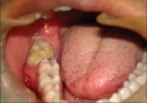
The formation of large sizes grows into the dental bone, thereby leading to its thorough destruction, the formation of ulcers and purulent fistula. It often develops without painful sensations, is characterized by gradual growth and absence of complaints from the patient.
Contents
- Causes of tumor development
- Current classification
- Symptom of the disease
- Diagnosis and treatment
- Prognosis and prevention of disease
Causes of tumor development
Most often, the tumor is detected accidentally - during radiography or in the procedure for tooth extraction. Sometimes, instead of a bone of the usual degree of density, a densified formation is found that can not be easily treated with conventional dental instruments.
There are several reasons for the development of tumor formation. It can result from an abnormality in the development of the dental embryo or surrounding tissue, genetic predisposition, trauma or infection of the oral cavity.
When studying the composition of the tumor, it reveals the remains of the epithelial islets of Malassa, the cells of the oral mucosa, enamel particles, pulp, dentin. In addition, the odontoma may contain a uniform dental tissue.
According to traditional medical concepts, odontomes are benign tumors. Nevertheless, in 4% of cases, there is a possibility of their degeneration into malignant ones. This process is especially prone to soft odontoma.
Modern classification
Depending on the reasons for the appearance of an odontoma: 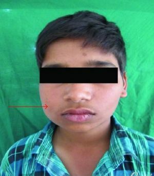
- is simple;
- complex;
- cyst.
A simple form of tumor formation is formed as a result of a violation of the development of one dental embryo, it is complete( formed from all tissues of the tooth) or incomplete( having in its composition one kind of solid dental substance).
Complex odontomas are formed by chaotic joints of teeth or tooth-like tissues. The development of this type of tumor is associated with disorders in the formation of several dental embryos. The X-ray pattern shows a tumor with prominent projections of rounded or irregular contours.
Complex odontomas are divided into compound and mixed. The composite complex odontoma contains several properly formed teeth, which have a deformed shape. Mixed consists of randomly arranged hard dental tissues.
The cystic form of education develops in the form of a cyst, in which the envelope contains a tumor. Around the formation, the presence of a transparent aureole is noted.
Depending on the structure, the tumors are divided into soft and hard.
Soft odontoma is less common than solid. It is often diagnosed among patients of an early age. The stage of the formation of permanent teeth is most "favorable" for the development of the tumor.
A tumor can develop in both the lower and upper jaws. It looks like an education consisting of gently fibrous connective tissues. Soft formations are considered precursors of the appearance of solid, are prone to relapse and transformation into malignant tumors.
Hard contains dental and periodontal tissues that are randomly distributed and have varying degrees of maturity. Hard odontomas are diagnosed several times more often soft.
Record composite odontoma for 232 teeth:
Symptomatic of the disease
 Symptomatology depends on the variety of odontoma. In most cases, it grows slowly and stops growing with the completion of the process of forming teeth. In the course of its development, complaints from the patient may not be forthcoming. As complications, inflammation foci in the localization of the neoplasm are noted.
Sometimes there is a constant growth of solid odontomas. A tumor of enormous size "settles" in soft tissues or in the periosteum. Its features are painless and a tendency to become infected, which in turn leads to the spread of the inflammatory process. The development of infection leads to an exacerbation, frequent occurrence of purulent fistula.
Solid odontomes are able to "push" and damage the embryos of teeth, as well as already matured teeth.
The tumor has an outline of an oval or an irregular prism. On the roentgenogram it looks like a dense body with uneven spine-like borders, often reminiscent of a raspberry berry. Education always surrounds the light portion in the form of a band( capsule)
Diagnosis and treatment of
The main diagnostic methods are clinical examination, X-ray examination and CT of the jaw. As an 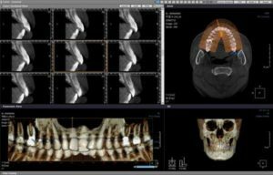 supplementary method, histological studies are performed to exclude the risk of a malignant tumor.
Treatment of such neoplasms requires a hospital stay and seeking help from the maxillofacial surgeons. Operative intervention is carried out under local anesthesia.
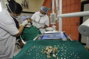
During the operation, the tumor is removed in stages, with small fragments. This is necessary in order to facilitate the removal process and prevent the possibility of fracture of the jawbone. After careful removal of the tumor, the wound is sutured. The 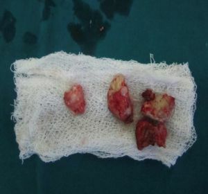 notch is filled with bone substance after surgical cleansing. In the presence of large tumors, partial excision of the jaw is carried out.
In case of significant destruction of the jaw bones and teeth, prosthesis is performed. An inalienable stage of treatment is further medical supervision of the patient's condition.
In developed countries, the treatment of odontoma is carried out using laser, cryogenic, ultrasound ablation, radiosurgery. Experienced specialists perform bone-plastic recovery operations at a high level.
Prognosis and prevention of the disease
Detection of tumors at the initial stage and timely access to professional medics contribute to a favorable prognosis of the outcome of the disease.
Do not delay the visit to the dentist when the initial signs of the disease appear. This will repeatedly worsen the situation and lead to an uncontrolled growth of the tumor. Together with this, the risk of degeneration of odontoma in malignant formation increases.
The basis of prevention should be regular visits to the dentist( at least twice a year), preventing the development of chronic infections and the effective elimination of inflammation. Detection of the disease at an early stage is a priority for modern dentistry and the medical industry as a whole.