
 Syndrome of the superior vena cava is characterized by the appearance of a severe circulatory disturbance in the basin of the superior vena cava.
The most common cause of this condition is lung cancer.
Before talking about the course of the syndrome of the inferior vena cava and about the methods of treatment, briefly describe what the hollow vein is.
Contents
- Anatomical approach
- Causes and risk factors
- Symptoms and signs of pathology
- Diagnostic methods
- How should I treat the disease?
- BASIC METHODS
- Drug Approach
- Surgery
- Disease Forecast
Anatomical Approach
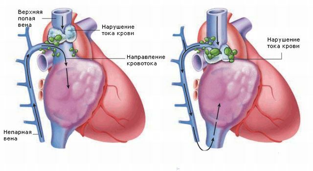
The superior hollow vein is a thin-walled vessel located in the middle mediastinum and surrounded by relatively dense structures such as the thorax, bronchi, trachea, and aorta. The vein is surrounded by a chain of lymph nodes all along its length.
In the inferior vena cava, the physiological pressure is low, which is a contributing factor for easy blockage of the vein with various lesions of the surrounding structures.
Upper hollow Vienna collects blood from the head, neck, upper chest, and also from the upper limbs. Compensatory function in the case of violation of the permeability of the veins is performed by anastomoses that connect the basin of the lower and upper hollow veins.
The unpaired vein is the main one. However, even despite the abundant amount of collaterals, they can not fully provide compensation for blood flow in the superior vena cava.
Therefore, the syndrome of compression of the superior vena cava is characterized by a pressure rise up to 200-500 mm water column.
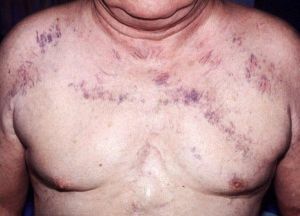
The photo shows the area that affects the syndrome of the superior vena cava.
Causes and Risk Factors of
The main mechanisms of the syndrome of suppression of the superior vena cava are:
- sprouting of the vena cava wall with a malignant neoplasm;
- thrombosis;
- compression from the outside.
After analyzing the data on the causes of this syndrome in different series, it was found that the most common cause is lung cancer, in 80% of cases the syndrome developed against the background of the right lung infection.
There are also non-tumor risk factors for the onset of syndrome:
- congested goiter;
- purulent mediastinitis;
- sarcoidosis;
- silicosis;
- constrictive pericarditis;
- post-radiation fibrosis;
- teratoma of the mediastinum;
- idiopathic mediastinal fibrosis;
- a long standing catheter in the channel of the hollow vein.
 To whom and in what cases is a phlebography prescribed. What rules for the preparation and conduct of the procedure exist.
 To give an answer to the question of Varifort cushion cheating or the truth, we have studied many reviews and materials on the topic and made an unambiguous conclusion.
Symptoms and signs of pathology
Approximately in two third patients, there are complaints of swelling of the face and neck, the occurrence of dyspnoea at rest, cough, sleep problems in the prone position, as the severity of these symptoms increases.
In one third of patients, the symptoms of upper vena cava syndrome manifest as wheezing, indicating laryngeal edema and the risk of obstructive airway obstruction. Increased pressure in the veins can lead to a swelling of the brain with the corresponding symptoms.
There is visible blood overflow of and as a result, edema of the face, collar zone, upper limbs, cyanosis of the skin and visible widening of the superficial veins.
In very rare cases, with the rapid development of blockage of the superior vena cava, there is a significant increase in intracranial pressure, cerebral edema, cerebral vascular thrombosis or even hemorrhagic stroke.
Diagnostic methods
The main diagnostic methods for the syndrome of the superior vena cava are: magnetic resonance imaging, computed tomography, phlebography. 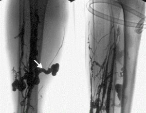
Additional instrumental studies are conducted to clarify the location and nature of blockage of the superior vena cava. The most informative in this regard are computed tomography and X-ray contrast angiography .
The results of these studies allow determining the localization, extent and nature of vein obstruction, collateral blood flow. You can determine the ratio of the tumor to other structures located in the mediastinum and thorax.
Accurately established tumor localization as a result of computed tomographic examination allows a safer sampling of the material to determine the morphological structure of the tumor.
How should I treat the disease?
The correct selection of the optimal therapy is based on the causes of the development of the syndrome of the inferior vena cava and the rate of progression of the process.
In half the cases the syndrome has time to develop more before the diagnosis. The determination of the initial process that caused this condition is the main component of effective therapy.
Only in the severe course of the pathological process with the threat of a patient's life, treatment is started without setting the main diagnosis.
Therapeutic measures in the syndrome of the superior vena cava are aimed at the relief of the symptoms of .In 50% of cases, the process is caused by a potentially curable disease.
Basic techniques of
For this purpose, in addition to , oxygen therapy and elevated position are sometimes performed by intubation, tracheostomy, and anticonvulsants .The patient is also prescribed diuretics and corticosteroids.
Radiotherapy is an effective method for treating upper vena cava syndrome in tumor diseases. Its efficiency reaches 70-90%.
Chest irradiation should be started as soon as possible. Emergency radiotherapy is indicated in the presence of respiratory failure or in the presence of symptoms from the central nervous system. Chemotherapy is used as the first line in the presence of a tumor that is sensitive to cytotoxic drugs.
Drug Approach
Therapy with anticoagulants or fibrinolytic drugs is directly used in vein thrombosis.
Assign these drugs only if a venous thrombosis is detected during phlebography or if there is no effect and signs of improvement from treatment in other ways.
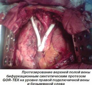
Surgery
The most promising surgical method for treatment of compression of the superior vena cava by a tumor or mediastinal fibrosis is the implementation of percutaneous endovascular balloon angioplasty with stent placement at the site of narrowing of the vein lumen.
Prognosis for
Disease Delayed results of therapy for upper vena cava syndrome are due to the underlying disease and the possibilities of its radical treatment. In acute syndrome, the patient may die.