
 Parks Weber Syndrome( congenital angiodysplasia) is a disease manifested by an increase in subcutaneous blood vessels.
The basis for the development of this syndrome is the presence of congenital defective anastomoses between the veins and arteries. The stage of severity of the disease depends on the number of anastomoses and their magnitude. Depending on the diameter, the anastomoses are divided into two types: microfistulas and macrofistulas.
The site of fistula localization is the zone of the popliteal, femoral and tibial arteries. Passage of arterial blood through arterio-venous fistulas can lead to an increase in blood pressure in the veins. High functional load leads to a change in the histological structure of the venous wall.
As a result, its miotic fascia is enlarged and an endogenous elastic membrane is formed. The presence of fistulas leads to the entry of arterial blood into the venous canal, causing oxygen starvation and disturbance of metabolic processes.
Causes and mechanisms of the disease
Most often, the disease occurs against the background of pathological changes that occur in the area of the location of the legs of the brain. These changes can be caused by a disorder of cerebral circulation, the formation of tumors and a violation of the integrity of the cerebral vessels.
The appearance of pathologies can also be associated with the pressure of tumor formations on the legs of the brain. In this case, the tumor can be located at a certain distance from the area of the legs.
The mechanism of the nucleation of Parks Weber syndrome consists of the following factors:
- subarachnoid hemorrhage in the middle cranial fossa;
- blood circulation disorder inside the main arterial vessel;
- development of inflammatory processes in the meninges in the brain base region;
- passage of pathological processes in the temporal lobe of the brain.
There are several risk factors in which angiodysplasia may occur:
- the presence of craniocerebral trauma;
- hemorrhagic or ischemic stroke;
- neoplasms in the brain are benign and malignant in nature.
Clinical picture of
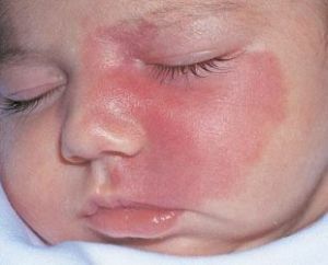
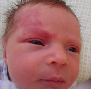
The first signs of the syndrome are manifested in the form of capillary ectasia and systolic-diastolic noise. At the same time, hypertrophy or stretching of limbs occurs, skin temperature rises and skin color changes( appearance of so-called wine spots).General changes in the body are manifested in the form of destruction of the main circulation.
The manifestation of all the above symptoms depends on the size and number of arteriovenous sastias and their location. The time of the course of the illness( fast or slow development) depends on the same factors. At the initial stage of the disease, the veins increase is insignificant.
With palpation, the convoluted veins are easily compressed, but after the pressure stops they appear back. In some cases, there is a pulsation of veins.
In the future, the affected limbs increase in length and volume. The lengthening of limbs by eight to ten centimeters is noted.
The cause of hypertrophy is the acute vascularization of bone growth zones and the buildup of blood pressure around the epiphyseal cartilage. The earliest and invariable sign of Parkes Weber's syndrome is an increase in skin temperature over the location of the fistula.
This symptom occurs due to arterial blood flow to the venous system and inhibition of venous outflow. The difference in skin temperature between the patient and a healthy limb is five to eight degrees.
Diagnostic criteria

In the photo, "wine spots" - a characteristic symptom of capillary angiodysplasia
To date, no specific diagnostic methods have been developed that could completely determine the presence of Weber's angiodysplasia.
Therefore, the diagnosis of this disease has certain difficulties.
For the diagnosis is used a full research complex using laboratory and instrumental diagnostic methods.
Laboratory diagnostics consists in applying methods of examining brain functions. In the presence of the disease, there are no changes in blood and urine composition, so taking such tests will not help to identify the disease.
In some cases, a spinal puncture is taken. In parallel with the puncture, the pressure of the CSF is determined. As for the used instrumental methods, they include the following research methods:
- Investigation of the vascular network of the fundus - detection of edema, vascular spasm, hemorrhage and vascular fullness.
- Computed tomography .It consists in resonance radiation or absorption by electromagnetic means.
- Neurosonography .Carrying out ultrasound of structural elements of the brain( ventricles, etc.).
In difficult situations, the diagnosis is established by determining the saturation of the venous blood with oxygen. A change in the venous pressure in the affected limbs is also monitored.
What does modern medicine offer?
Very often, with Parkes-Weber syndrome, a conservative treatment is prescribed, which is the use of third-class compression knitwear. Recommendations for the use of laundry are given based on the symptoms of the disease. 
Laser removal of so-called "wine spots" is applied. In severe cases, a surgical procedure is performed, consisting in excising or ligating the anastomosing branches and dilated vessels.
Patients whose changes in the extremities are large enough and their functions are completely lost, their amputation is carried out.
The prognosis for this disease is unfavorable. This is due to the gradual development of cardiac deficiency. Of the complications, there is a possibility of the development of trophic disorders in the tissues of the extremities. With this syndrome, there is a predisposition to the formation of blood clots in the vascular wall. In case of injuries, internal bleeding is possible.