
 Ileofemoral thrombosis is an pathological process characterized by occlusion of the vein lumen at the level of the iliac and femoral vessels by a thrombus.
The thrombus obstructs the blood flow to the lower parts of the leg, resulting in clinical manifestations of the disease. More often Ileofemoral thrombosis affects elderly patients who have cardiovascular diseases, diabetes, obesity, just the elderly and cancer patients.
Ileofemoral thrombosis of the lower extremities often appears with severe injuries in , in traumatic and prolonged operations, in pregnant women before and after childbirth.
It can also complicate emerging infectious and purulent diseases. All of the above conditions are risk factors for thrombotic complications.
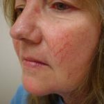 How is treatment of couperose on the face with a laser, are there any contraindications and complications after the procedure?
You need to know this if you have prescribed Anavenol medicine - instructions for use, indications and contraindications, reviews of doctors and patients, and other useful information.
Content
- Causes of
- disease clinical picture
- Diagnostic techniques
- Treatments
- Surgery
- Complications thrombosis
- Prevention Tips
- Conclusions
Causes of
disease in developing ileofemoralnogo thrombosis very important wins back change in the internal wall of the vessel at the affected limbs. Because of its damage, the release of substances responsible for the inflammatory process and activating the platelets.
All this contributes to the activation of platelets and the start of blood clotting. The site of damage to the inner wall of the affected vessel becomes a place of platelet aggregation, which is why a thrombus is formed.
The aforementioned cause in combination with certain factors and becomes the cause of ileofemoral thrombosis.
Such a situation can occur in such situations:
- For example, in operation , when a person lies motionless, with the lower limbs pressed to the operating table, blood stagnation occurs in the bloodstream, which contributes to the formation of a thrombus.
- Prolonged bed rest also promotes the appearance of blood stagnation in the lower limbs. In injuries, the vessel wall is damaged, which is a provoking factor of thrombosis.
- Against the backdrop of infectious diseases and often the walls of blood vessels are affected by pathogenic microorganisms. It is believed that contraceptive
 funds can contribute to the development of thrombosis.
 - In humans, with oncological diseases , there is an increase in the activity of the blood coagulation system, which increases the risk of thrombus formation.
Possible congenital or acquired diseases of the blood system, such as thrombophilia. All these factors alone or in combination cause the formation of a thrombus with subsequent blockage of the major vessels of the lower limb.
Clinical picture of
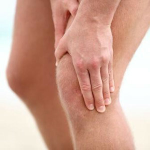
In the case of Ileofemoral thrombosis , the patient is concerned about the pain , localized on the antero-internal surface of the thigh, in the calf muscles, sometimes in the groin area.
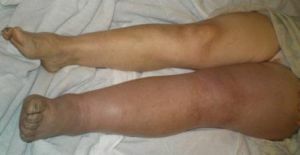
The extremity increases in volume and the edema spreads from the foot to the groin, sometimes with the transition to the buttock. The color of the limb varies from pale to cyanotic. When palpation along the trunk veins and in the groin, soreness is determined.
Sometimes the disease occurs suddenly, begins with acute pulsating pain in the limb, its cooling and numbness.
There is a rapid increase in edema, the movement of the toes becomes limited, there is a decrease in the sensitivity and skin temperature of the lower limb, weakening or completely disappearing pulsation of the arteries of the foot.
This form of ileofemoral thrombosis is called " pseudoembolic", or white painful phlegmation of , its occurrence is associated with a combination of deep vein thrombosis with severe arterial spasm of the diseased limb.
When a widespread thrombosis of all deep veins of the lower limb and pelvis occurs, a sharp increase in the limb in the volume occurs, it becomes swollen and dense.
The skin is characterized by a purple or almost black color. Bubbles filled with liquid appear on it. This clinical form is called blue pain reflux.
It is characterized by the emergence of strong tearing pains, absence of pulsation of peripheral arteries.
There are two stages of the flow of ileofemoral thrombosis:
- Prodromal stage. Characterized by pains of different localization, aching or blunt character, increased temperature.
- Stage of severe clinical manifestations. Characterized by the emergence of a classic triad of symptoms: color, swelling and pain.
Diagnostic procedures
Diagnosis of ileofemoral thrombosis is based on the clinical picture of the disease and data of instrumental research methods.
The simplest and safest method for detecting thrombosis, and quite effective, is ultrasound duplex scanning .
Using this type of study, you can see the lumen of the vessel, clarify the degree of reduction in the lumen of the vein, and also to determine the extent of the thrombus and its mobility.
With complete blockage of the lumen of the blood vessel with a thrombus, there will be no blood flow, and if there is partial obstruction, the thrombus will flow through the narrow, remaining sections of the vein lumen.
Duplex scanning is used for the differential diagnosis of ileofemoral venous thrombosis with edema of the lower extremities arising from other diseases.
Also used for diagnosis of thrombosis is the phlebography .This method of investigation consists in the use of X-ray equipment to determine the presence of a thrombus in the lumen of the vessel.
Carry out this procedure with the introduction of a special contrast in the vessel, which will evaluate the movement of blood. Sometimes, in difficult cases for differential diagnosis, magnetic resonance phlebography is used.
Treatment procedures
Treatment of ileofemoral thrombosis, as well as other deep vein thrombosis, is performed in a hospital.
Usually uses conservative therapy with , much less often surgery. For most patients, heparin is the drug of choice in the treatment of 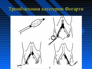 ileofemoral thrombosis.
There is a special course of treatment with heparin, the high effectiveness of which has been proved by numerous clinical studies in many medical centers.
Conservative treatment is carried out in combination with early activation of patients. It is advisable to lift the foot end of the bed at an angle of 15-20 degrees.
Patients are shown to adhere to bed rest only at the initial stage of the disease in case of pain and edema of the affected limb.
When stihanii pain and reduce edema, it is advisable to designate a set of special gymnastic exercises that contribute to improving venous outflow. These exercises are necessarily controlled by the methodologist of the cabinet of physiotherapy.
Operative intervention
The operative methods of treatment of include:
- thrombus removal with Fogarty catheter;
- bypass operation.
Complications of thrombosis
The most frequent and formidable complication of any thrombosis of the veins of the lower limb, including ileofemoral thrombosis, is pulmonary embolism of the pulmonary artery.
With incorrect treatment of thrombosis, 50% of patients have a risk of pulmonary embolism during a three-month period. For its prevention, special cava filters in the inferior vena cava are often installed.
Their shape resembles an umbrella, in which there are special holes for the passage of blood.
Correctly selected and performed treatment of acute deep vein thrombosis of lower extremities with the use of anticoagulants helps to reduce the risk of thrombus spread and pulmonary artery embolism up to 5% and lower.
Advice on prevention of
Prevention of ileofemoral thrombosis is very important, as it allows patients to get rid of life-threatening complications of this disease.
 The extent of the need for prophylaxis for thrombosis is particularly high in patients at increased risk. These include elderly people, patients with cancer and severe cardiovascular diseases, people suffering from obesity.
Preventive measures are usually directed to to prevent the occurrence of venous congestion, by accelerating blood flow. This is achieved by bandaging the legs with elastic bandages.
When the superficial veins are squeezed with bandages, the blood flow in the deep veins is accelerated, thus inhibiting the formation of thrombi. An adequate mode of activity is also recommended.
Conclusions
Ileofemoral thrombosis is a very serious threat to human health, but is treatable, especially timely. So you can not give up. You need to see a doctor and follow his treatment recommendations.