
 This phenomenon, like adentia, which is the absence of teeth on both the upper and lower jaws, is quite often found not only in older people, but also in the young population.
This pathology should be eliminated as soon as possible. This is due not only to purely aesthetic moments, but also to the further development of serious complications.
In order to choose the most effective method of treatment, the dentist must first thoroughly study the features of the jaw structure of a particular patient and classify it according to existing rules.
With the help of standard methods of qualification, it is easy to choose the right strategy for treating a patient, and also to facilitate the work of dental technicians from the point of view of manufacturing prostheses. Also, it will make it possible to minimize the likelihood of any complications and problems at each stage of treatment.
Contents of
- Types and features of toothless jaws
- Types and features of toothless jaws
- Classification by Schroeder
- Classification by Keller
- Classification by Oksman
- Classification by Courland
- Classification by Dojnikov
- Types and features of toothless jaws
- Process for creating impressions
- Mucous membrane of prosthesis box
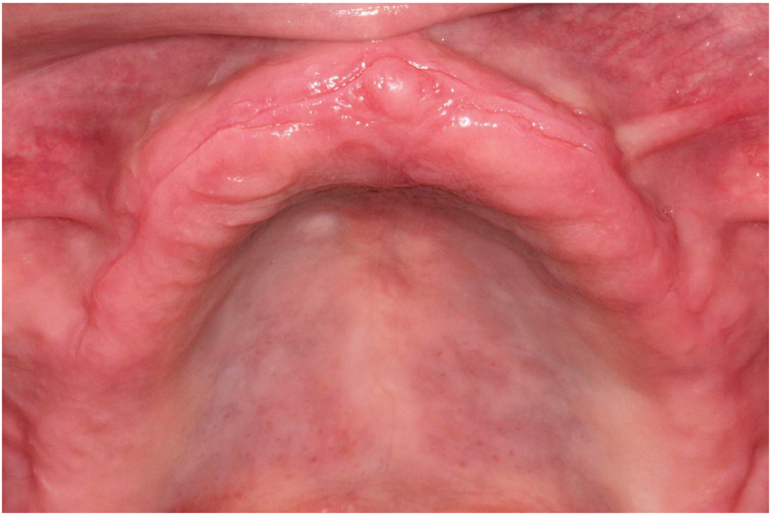
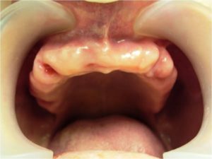
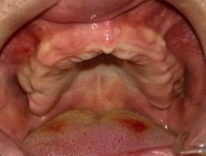
Types and features of edentulous jaws
There is no single standardized classifier in modern medicine. This is due to the fact that there are many transitional variants between all known types of jaws, which complicates the creation of a single classification. At the moment, several of the most well-known classifications are used.
Schroeder classification
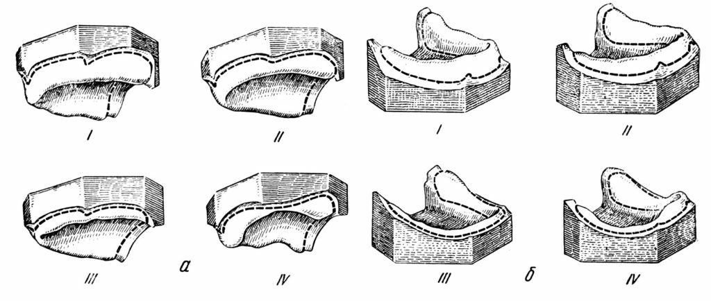
So, the Schroeder classification distinguishes three types of upper jaws with missing teeth. These types differ in the degree of depletion of bone tissue in the region of the alveoli:
- First .This is a frivolous degradation of the gear part. With such a clinical picture, the jawbone tubercles and the areas of the upper dentition that serve as a dentition for the teeth are clearly pronounced. Thus the sky differs good depth. The bends of the mucous membrane and the area of fixation of the muscles are located quite at a decent height. Physicians consider this type of structure the most suitable for the manufacture and subsequent installation of the prosthesis. This is due to the fact that in this case there are no physiological abnormalities that would directly prevent the installation of the teeth.
-
 Second .It is determined by specialists on the basis of the average severity of alveolar axon degradation with its fuzzy expression. In such patients, physicians observe a moderate depth of the palatal plane. In this case, the transition fold is displaced closer to the alveolar ridge. When fixing the jaw prosthesis in the presence of such a clinical picture, the probability of deterioration in the quality of its fixation increases. Such problems arise against the background of muscle overstrain that are involved in mimic movements.
 - The third .You can observe a serious destruction of the bones at the base of the jaw. Completely smooth out all the mounds and crests of the alveolar. The sky acquires a flat form. At the mucous membrane the fold is very low directly in the same plane as the sky itself. This form causes the greatest problems when carrying out dental prosthetics. This is due to increased mobility of structural elements due to the pathological structure.
Keller classification
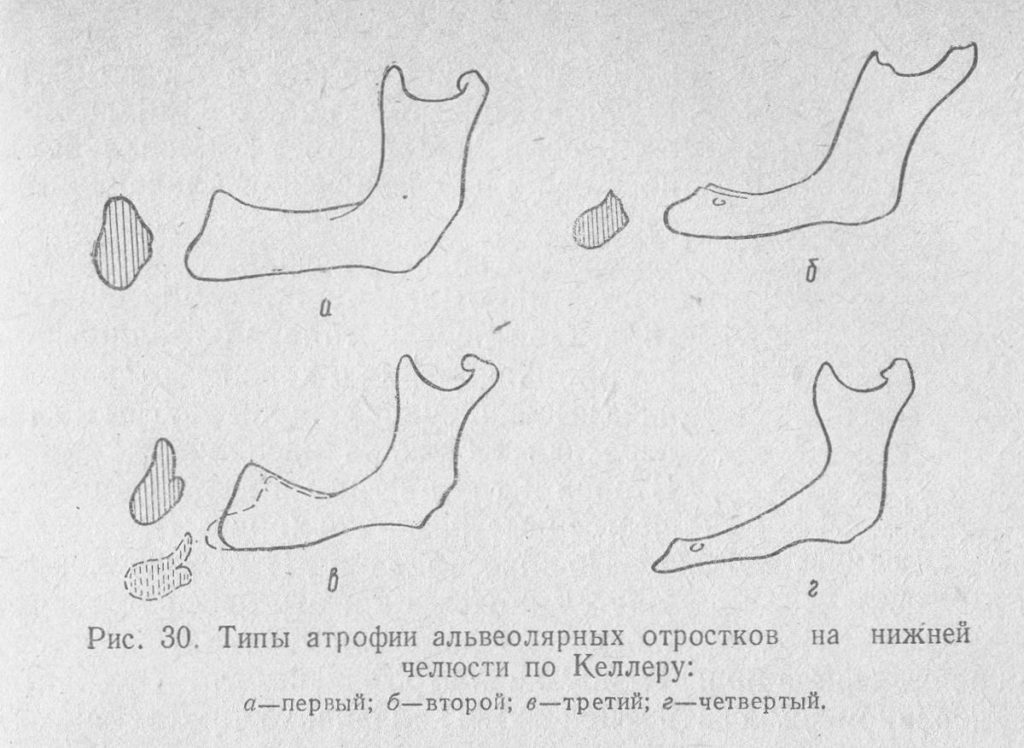
To somewhat simplify the process of restoring parts of the lower row of jaws, Keller's classifier was specially created. In this classification there are four types, namely:
- First .It is a minor jaw bone atrophy and a slight smoothing of the alveolar elements. This type is ideal for manipulating the installation of prostheses. The folds of the membrane, as well as the muscles, are attached in the region of the beginning of the alveolar ridge. As dentists note, this type is extremely rare among patients. Most often, such a jaw is a consequence of the simultaneous removal of all teeth during a short period of their absence.
- Second .It is an appreciable process of tissue destruction. On the general background of the base of the mouth, the crest is slightly prominent. Moreover, it has a relatively sharp surface, which seriously complicates the reliable fixation of the denture. In this case, the muscles have attachment in the region of the alveolar ridge. Some nuances of the structure of this jaw can cause some discomfort and even painful sensations in the patient when using a prosthesis.
- Third .It is allocated by dentists in those patients who have an early extraction of teeth, which are located on the sides. This type is characterized by thinning of the appendages of the alveoli in the region of both molars and premolars. At the same time, the total volume of bone tissue in the central section is preserved. In this case, dental prosthetics are allowed as in the lateral part of the row of teeth there is a flat surface, which is excellent for reliable fixation of artificially created molars. It is also worth noting that due to the preservation of the hillock of the alveoli in the central part, the possibility of slipping of the prosthesis during chewing of solid food is excluded.
- Fourth .Characterized by a strong atrophic process of the alveolar zone in the area where the frontal incisors are located. Simultaneously with this side of the dentition, good tissue preservation is observed. In this case, the prosthesis is not fixed very well because there is a high probability that it can move and lose its stability.
Oksman's classification
Outstanding figure in Soviet medicine, Doctor of Science, Professor Oxman has developed his own system for determining the type of jaws without teeth.
According to Oksman, the upper toothless jaw can be conditionally divided into the following four types:
- First .In determining the first form, a man has a long alveolar axon and pronounced knolls. In such a clinical picture, the palatine surface is bright. Muscles in this case are fastened at a sufficient height.
- Second .It differs by more noticeable thinning of bone tissue with uniform atrophy. In comparison with the first type, the sky becomes less deep. In the center of the alveolar sector, the membrane of the mouth is attached.
- Third .In the case of diagnosis of the third type, a significant and uniform atrophy of the upper jaw occurs in a person. The human sky eventually becomes completely flat, and the shell is attached to the crest.
- Fourth .If it is a question of the fourth type, then in this case there is an uneven atrophic process of the areas of the alveolar. In general, all pathological changes affecting the jaw fully correspond to those described in the three previous types.
As for the classification of the lower jaw, Oxman, based on the stages of the atrophic process of bone tissue and certain anatomical changes, identified the following four varieties:
- The first form of .In the alveolar process a considerable height is observed with a simultaneous low position of the mucosal supports and all frenums.
- The second form is .There is a uniform process of changing the density of alveolar tissues at their average level of severity.
- The third form is .The alveolar segment is weakly expressed or completely absent. Deformation is often observed.
- Fourth form of .There is an uneven depletion of bone tissue in different parts of it. This is dictated by different times of tooth loss.
Classification according to Courland
According to the systematization developed by Dr. Kurlyandsky, there are four separate classes of jaws without teeth:
- The first group of .The first group includes patients who can observe the alveolar process, which extends beyond the place where the muscles attach.
- The second group is .Combines the jaws with the thinning of bone tissue in the region of the jaw bone with its location on one level with the place of attachment of the muscles.
- The third group is .The patient has a serious atrophy of those parts that are located below the level of the site of attachment of muscles. The fourth group is .It assumes that the bone in the places where premolars and molars were previously located, is seriously depleted. The fifth group is .Atrophic processes completely affect the tissues in those places where the front teeth were previously placed.
Doinikov classification
Doinikov's classification system for toothless jaws is similar in many respects to the classifier proposed by Schroeder. However, it has some significant differences, based on the features of thinning of individual parts of bone tissue:
- The first form of .Both jaws have pronounced crests and alveolar processes. On the plane of the palate, the oral mucosa is uniformly distributed. At the same time, it has good compliance. The folds are located a short distance from the top of the ridge.
- The second form is .All patients have an average level of destruction of the dental tubercles. The total depth of the sky decreases in comparison with the first form. The torus is rather well expressed.
- The third form is .It is impossible to trace the alveolar parts of a row of teeth. Bugry and body seriously decrease in contrast to the parameters of the normal state of the jaw. The sky acquires a completely flat form with a fairly wide torus.
- Fourth form of .Only in front one can observe a pronounced alveolar process. On the side, the sites are seriously atrophied.
- The fifth form is .Atrophy is observed in the anterior part, while maintaining the density of bone tissue on the sides.
Process of creating prints
With the help of creating impressions, it is possible to form both diagnostic and fully working forms, which are subsequently used for subsequent casting of denture designs. For today in stomatology it is accepted to use some basic kinds of impressions.
 Anatomical type of impressions can be removed using conventional spoons for impressions and use of dental gypsum.
These prints have high edges. In this case, it is not customary to use functional tests. Because of this, it is impossible to take into account the general condition of the oral tissues, which directly border on the prosthetic bed.
The functional type of impressions is made using an individual spoon and a functional test, which makes it possible to determine the condition and overall level of the possibility of movement of the mucosal folds. Unlike the previous type of impression, in this case the edge of the impression is located slightly lower. At the same time, the border of the finished prosthesis affects the shell no more than 2 millimeters.
Functional dental prints, in terms of pressure on the mucosa, are divided into three separate types:
- Discharge impression type .It is removed with the help of gypsum while minimizing the pressure on the shell. Compression prints .Used only in case of good compliance from the mucosa. They are made by using silicone, gypsum or thermoplastic paste and with a little pressure.
- Combined class of prints .Allows you to squeeze those parts of the mucosa that are good compliant. In this case, areas with poor compliance are not overloaded.
The mucosa of the prosthesis box
In addition to the accessory of the toothless jaw to this or that variety, the specialists take into account the features and characteristics of the mucosa before the prosthesis, which is directly located in the prosthesis bed.
It is customary to distinguish three main types of mucosa:
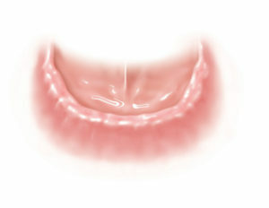
- Normal .Normal is characterized by good ductility and good hydration. Most often, this mucosa has a gently pink tinge. This type is best suited for manipulating dental prosthetics.
-
 Hypertrophied .For the hypertrophied, a loose structure is characteristic, as well as the presence of some intermediate structures. She has good moisturizing. In this case, due to high compliance, it is often possible to observe high mobility of the installed dental prosthesis.
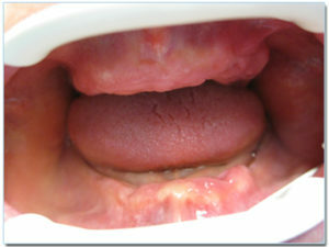
 - Atrophied .The atrophied shell has an increased structural density with a simultaneous poor moistening. Often it has a white tint. Part of the mucosa located on the maxillary process is attached to the periosteum. In this case, it is more difficult for specialists to perform a qualitative denture due to the specificity of the mucosa.
With prolonged absence of teeth in the tissues of the bones of the dentition and the oral cavity serious pathological processes begin to occur:
- atrophy of bone tissues;
- complete destruction of the mucosa in the mouth;
- functional changes in the jaw joints;
- the onset of inflammatory pathological processes;
- problems with full nutrition;
- problems with speech;
- disruption of facial structure due to exhaustion of facial muscles.
Most doctors agree that one should not postpone for a while with dental prosthetics in the absence of teeth.