
 Adamanthoma( another name - ameloblastoma) is a tumor of an odontogenic type. Despite the fact that the new formation itself is of a benign nature, its malignancy is possible.
To avoid a negative prognosis, timely and correct treatment is necessary.
Contents
- Overview
- Causes and pathogenesis
- Classification of
- Features of the
- Clinic Diagnostic approach
- Surgical care
- Rehabilitation period
- What does the prediction of
depend on General information
Adamantoma is formed from the epithelial tissue of the dental papilla or enamel cells( involved in tooth development).The site of tumor localization is the embryonic layer in which tooth enamel is produced. There can be a pathology in any area of the jawbone.
In 90% of cases, the neoplasm is diagnosed on the lower jaw in the area of the molars( more often near molars and premolars).In 10% of patients, ameloblastoma is detected on the upper jaw. There is a disease in people between the ages of 20 and 50 years.
The pathology develops slowly, in some cases the disease manifests itself only after several years. Education, increasing in size, repels the nearby bone, which begins to stretch. When the tumor grows to such a size that the dental bone is destroyed, then nearby soft tissues are affected.
Dangerous for complications. The most unfavorable prognosis is the development of cancer. Similarly, with ameloblastoma, a pathological process can spread in the sinuses of the nose, then into the cranial base and the brain.
Causes and pathogenesis of
The reasons for the progression of the tumor are not defined in medicine. However, specialists suggest that 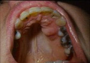 adamanthinum, possibly, is a consequence of improper formation of the teeth rudiments. Also, some experts believe that the disease is the result of a pathological condition of epithelial odontogenic remains in the jaw.
These reasons are a consequence of already established deviations in the physiological state of the jaw joint. The provoking factors, which are a peculiar impetus for the disease, are:
- injuring the jaw or oral cavity;
- infectious inflammation of the gums or teeth;
- viral infections.
Modern medicine suggests that the development of education is possibly affected by the lack of a number of minerals and proteins.
Classification of
Adamantina is divided into two forms, each of which has its own developmental features. Also, five types of tumors are distinguished depending on the characteristics of the microscopic characteristics.
Tumor forms:
- With , massive form, one or more cysts are merged into one formation. In this case, a spongy tissue is formed inside the neoplasm. On X-ray, a solid form can not be determined.
- The cystic form is most common. In this disease, the patient develops separate or connected partial neoplasms, within which there is a thin epithelial tissue.
Ameloblastoma can have a different structure, so there are five types of pathology:
In the photo, the follicular ameloblastoma of the mandible in the advanced stage
- is basally celled;
- follicular;
- akantomatous;
- granular-checkered;
- pleomorphic.
The most commonly diagnosed follicular and pleomorphic type.
However, with the development of even one neoplasm, two different types can be combined in it.
Features of the
clinic It is difficult to detect ameloblastoma, since before the appearance of external defects the tumor can progress asymptomatically.
The most characteristic features are:
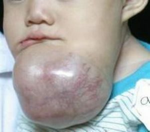
- asymmetry of the face of different degrees;
- pain, similar to toothache;
- displacement and loosening of the teeth;
- increasing deformation of the jaw;
- sound crunch when jaw motion.
For a long time, the skin in the affected area remains unchanged. Later, the pathological process spreads, accompanied by inflammation of the oral cavity( fistula formation with purulent discharge).When examining the jaw( palpation), a bumpy or smooth part of the bone can be felt, which has a swelling, springing and deforming when pressed.
In 90% of cases, ameloblastoma forms on the lower jaw, while the new formation itself passes into the maxillary sinus and may not have visual deformation for a long time. Moreover, the maxillary type of the disease, which is diagnosed more easily, often causes complications due to the risk of spreading into the sinuses of the nose, orbit and brain.
Diagnostic approach
It is extremely rare to detect the disease before the appearance of visual symptoms. As a rule, this happens by chance in the treatment of teeth. Diagnosis includes a number of studies, on the basis of which the diagnosis is confirmed.
Comprehensive study includes:
- computed tomography;
- X-ray examination;
- cytology.
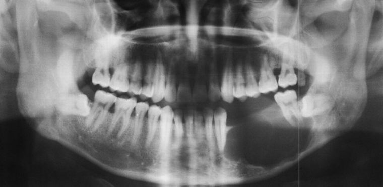
A dark spot on the x-ray reveals the tumor
Mandatory are the studies of blood and urine, as well as the delivery of blood to the oncomarker to determine the risk of malignancy of the tumor.
Differential diagnosis is also performed to confirm the disease, which can be confused with the development:
- dental cyst;
- squamous cell carcinoma;
- carcinoma( cystadenoid) salivary gland.
The most reliable methods of examination for the diagnostic purposes of this education are x-rays and cytology. Based on the general diagnostic results, the type of treatment and further prognosis are determined.
Surgical care
It is possible to get rid of progressive adamanthamum only surgically. Therapy is used only as a method of additional impact on the pathological process. The treatment regimen is always selected individually, depending on the prevalence of inflammation, the site of localization and the stage of the disease.
Modern diagnostic methods can determine the disease before the tumor reaches a large size. The operation can be carried out in several ways.
Methods of tumor removal:
- without resection;
- with resection;
- with partial jaw exarticulation.
In surgical intervention without resection, the jaw joint does not move, due to which its functionality is not violated. The patient's face after surgery has no external defects. However, this kind of surgical intervention is only permissible if the disease is diagnosed at an early stage.
If ameloblastoma has acquired significant dimensions and spread to other areas, the surgeon resection or resorts to partial eccentricity( removal of part of the jaw bone).
Neither the first, nor the second type of surgery does not exclude relapse, because the enlarged tumor can not be completely removed completely.
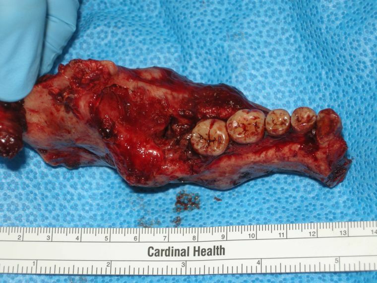
Removed part of jaw affected by adamantine
Rehabilitation period
Treatment of ameloblastoma is not possible without tissue damage and temporary( and sometimes permanent) dysfunction of the jaw. To restore the patient requires a rehabilitation period, which can last from several weeks to several months.
After the operation, therapy is mandatory:
- antibacterial;
- corrective;
- is symptomatic.
The action of drugs prescribed by a doctor is aimed at preventing and preventing the inflammatory process, reducing the risk of relapse.
Also, the patient must adhere to a special diet that excludes the use of hard and rough food, which is difficult to survive. In this case, after each meal, it is necessary to rinse the oral cavity to prevent the ingestion of food residues into the operated cavity.
 If the operation has led to deformation of the jaw, corrective therapy is used to restore the shape, consisting in the establishment of a special orthodontic construction or bone plate.
Full complex rehabilitation allows the patient to recover not only physiological, but also psychologically. If the operation has led to deformation of the person, the patient must necessarily visit the psychologist.
What determines the prognosis of
The consequences of a tumor depend directly on the stage at which it was detected. The earlier the disease is determined, the less negative the consequences. With a timely diagnosis, the forecast is quite good. But it should be remembered that a complete lack of treatment can lead to cancer.
Preventive measures for the prevention of pathology do not exist, since the causes of the disease are not established, and most likely, are intrinsic.
Specialists advise regularly( at least twice a year) to visit the dental office, so that when there is adamanthinoma, there is an opportunity for early diagnosis.