
 The oral cavity is constantly exposed to various pathological processes, one of the most common tumor diseases is cementoma.
Tumor can develop in both adults and children. Characterized by cementom, as a benign formation, forming in the circumcision area of the tooth.
Contents
General
General
General description of
Among various tumors of the oral cavity cementum is considered one of the most dangerous, as in the process of development it does not reveal itself at all, and the formation gradually destroys the bone and epithelial tissue in the mouth.
Such, at first glance, a strange name, is associated with the peculiarity of the tumor that forms a tissue similar to the cement of the tooth. There are several types of pathology that have their own characteristics. Diagnosis of the disease in most cases in people between the ages of ten and twenty years, but it is possible to develop a tumor at any other age.
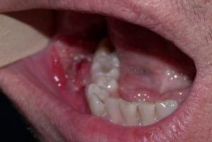
The tumor is localized mainly in the area of incisors and premolars of the lower jaw composition. External signs in the form of deformation of bone tissue are noted only if the growth of the tumor is considerable.
Modern classification of
Modern medicine identifies two main types of disease that are characterized by the area of the lesion:

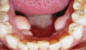
In the photo, the giant lower limb cementum
- local - the tumor occurs near the root of only one tooth;
- diffuse - pathology affects the area of several tooth roots, as well as affects bone tissue.
Depending on the specific type of cementum, the pathological process has specific causes and symptoms:
- Fibroma .The symptomatology of fibrinous cementoma is unclear. The tumor damages the bony cortical layer and forms mineralizing zones. The capsule is located on the outside.
- True .Signs of pathology are weak. As well as fibroma has areas of calcium accumulation. Neoplasm is covered with a shell due to what the pathological process has a limited area. Formed from the outside.
- Fibrous .Another name is periapical cement dysplasia. It is localized in the posterior part of the mandibular structure. For fibrous tumor characterized by the formation of a number of foci, bone destruction and places of accumulation of minerals.
- Giantformed cement. The most complex type of disease, in which there is a transformation into cement connective tissue. Neoplasm densely grows to the tooth root. The tumor has a high density.
Provoking causes of
Factors affecting pathogenesis are not defined even by modern scientists and physicians. Under the assumptions of scientists, the reasons are in a dysplastic or deterministic process.
It is possible that the disease is hereditary, since, for example, a giant cementoma often has a hereditary character.
Presumably, the following factors have a negative effect on the onset of development of the pathological process:
- permanent inflammatory processes occurring in the oral cavity: actinomycosis, periodontitis, sinusitis, etc.;
- regular injury: damage to the mucous caries teeth located in the mouth or when installing foreign objects( fillings, dentures, braces, etc.);
- single injury: stroke, bruise, mechanical injury to the gums or jaw bones.
Some experts also believe that smoking, radiation and other external factors of a similar nature can have a negative impact.
Features of clinical picture
 Symptoms of cementum, as already mentioned, arise only when the pathological neoplasm begins to increase significantly in its size.
Until this point, painful symptoms or visual deformity, as a rule, are not observed.
In advanced cases, the patient exhibits the following symptoms:
- a feeling of discomfort in the area, damaged dental tissue;
- pain when chewing food;
- pain during palpation;
- discoloration of tooth enamel.
If the tumor is overgrown, then external facial defects can be noted, and in some cases there is a gradual rupture of the mucosa and the epithelium of the oral cavity in the area of localization of the tumor.
Diagnostic methods
It is possible to determine the presence of cementoma only with the help of a special X-ray performed on the area of the jaw composition. It is enough 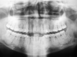 is often the diagnosis of dental diseases allows timely detection of pathology.
On X-ray images, the tumor manifests itself in the form of a darkened spot of a rounded shape localized in the region of the tooth root. In this neoplasm is so closely located to the teeth, which is one with them. Difference from other pathologies is the presence of dense sites with mineral formations. Similarly, bone tissue itself can undergo mineralization.
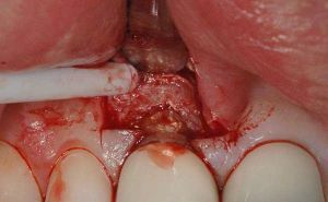
Once the disease is determined, treatment is administered immediately. Therapeutic methods to combat this pathological process can be used for certain types of disease. The main method of treatment is an operation.
Features of operation intervention
The operation is appointed by the attending physician. The actions of surgeons consist in the removal of the formed tumor with the use of resection. During the operation, the part of the bone that was damaged is removed. Similarly, the tooth that has been subjected to the pathological process undergoes removal.
The operation is necessary because of the high risk of complications in the form of spread of tumor formation in the upper jaw, damage to the integrity of bone tissue, as well as the occurrence of perforation on the oral mucosa.
 Do not remove the tumor in fibrotic and gigantiform type of disease. With these types of cementomas, surgical intervention can be avoided, since active growth is not characteristic of these tumor forms.
During the therapy, the patient is recommended to take anti-inflammatory drugs and have constant monitoring by the doctor. If complications arise, surgery can be prescribed, if the process of rejection of pathological tissues proceeds without a negative prognosis, then gradually the tumor disappears naturally.
To accelerate the process, specialists can perform mini-operations to create a saucer-like cavity, thereby restoring faster.
Consequences and complications of
Cementoma is a tumor formation that, although benign, can have serious consequences. Complications, as a rule, arise in neglected cases, or when the pathology is localized in the upper region of the jaw. 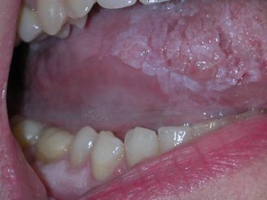
This is due to the risk of damage to the nasopharynx or nasal sinuses. The defeat of tissues can occur during surgery, or with the growth of the tumor.
The danger is also the output of pathological formation on the external side of the gum. Perforation can lead to the formation of holes from the bone to the mucosa. As a result, the infection begins to progress in the oral cavity, which can have a large area of spread.
What about prevention?
The reasons for the appearance of cement are not defined, therefore it is difficult to prevent the disease. However, if you consider factors that can provoke a pathological process, then you can reduce the likelihood of the disease.
In case of damage to the jaw or inner cavity of the mouth, you should immediately contact a specialist for assistance. It is also recommended to perform preventive examinations at the dentist, in time to conduct treatment for diseases of teeth and gums and properly care for the oral cavity.
When thinking about the need for prevention, it should be remembered that the disease can lead to the occurrence of concomitant pathologies and irreversible consequences in the form of deformation of the jaw, which will not remain invisible in the external appearance of the patient's face.