
 Sialografiya is an X-ray examination of the ducts of large and small salivary glands.
This radiopaque method is effective for detecting inflammation of the salivary glands, sialolitis, sialosis and other disorders.
Contents
- Methods of conducting the study
- Scope and contraindications
- How the
- is being carried out Decoding of the results
- Price question
Methods for conducting the
study There are several varieties of the sialogram:
- Thermosialogram .The essence of this research is the absorption of infrared rays by tissues of various structures. Thus, it is possible to measure the temperature in the salivary gland zone. To perform the diagnosis, a thermal imager is used, which helps to recreate a thermal map of the temperature of the face and neck. The increased temperature of the skin above the salivary glands indicates the presence of malignant neoplasms or the course of inflammatory processes in them. This method of diagnostics is absolutely safe and has no limitations to the application.
- Sialosonography .This is an ultrasound diagnosis, during which the tissues of the ducts absorb ultrasound. On the basis of this method, it is possible to determine the shape, parameters and correlation of duct tissues. Diagnosis also helps to identify sclerotic changes, the presence of sialites and the edge of growths.
- Pantomosialografiya .It is carried out in parallel with the contrasting salivary, two submandibular and two parotid glands. Diagnostic results are displayed in panoramic tomography. Due to the examination of the paired glands, even invisible inflammatory processes can be detected there.
- Digital sialogram .This is a method of investigation, which is carried out on specialized equipment with numerical data( angiography or X-ray equipment with a digital set-top box).In the course of this study, it is possible to conduct an analysis of the filling of the ducts and the emergence of a contrast medium from them.
- Sialadenolymphography .The lymphatic system of the salivary gland is analyzed for the diagnosis of diseases. During the diagnosis, a contrast agent is injected into the parotid gland, followed by serial sialadenolymphography. In the presence of growths, the filling defect is traced.
- Computerized sialotomography .A scan is made from the level of the hyoid bone and to the parotid glands. The resulting transverse section is displayed on computer tomographs. This method is used to diagnose tumors of salivary glands and salivary stone disease.
Scope and contraindications
Indications for the purpose of this study:
- presence of stones in the outflow ducts( salivary stone disease);
- sialolithiasis;
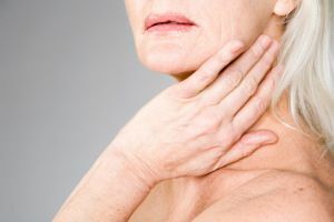
- presence of mumps( persistent inflammatory diseases of the salivary glands);
- presence of salivary gland tumors;
- definition of cysts of varying degrees of difficulty;
- definition of duct obstruction;
Contraindications to diagnosis:
- presence of acute inflammation of the oral mucosa;
- inflammation of the papilla of the duct of salivary glands( relative contraindication);
- high susceptibility of the body to iodine;
- pregnancy period( except for life threatening events).
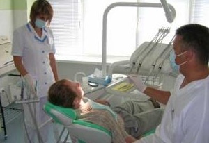
How the
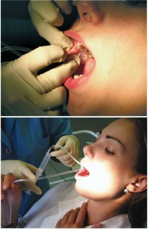
 is being conducted The essence of the sialogram is the introduction of contrasting substances into the salivary gland duct. This manipulation is carried out using a syringe.
Initially, contrast media are heated to a temperature of about forty degrees, so as not to cause cramps in the ducts. The volume of the required substance is determined based on the type of gland, age and sex of the patient. On average, the amount of the drug for the parotid gland is 2.5 ml, and for the submaxillary gland is 1.5 ml.
After this, the ducts are washed with the isotonic essence of sodium chloride and bougie with a subtle buzhem. A catheter with a contrast agent moves into the interior of the duct( about 3 cm) and is fixed on the skin of the cheek.
The saturation level of the ducts is regulated by performing a television X-ray inspection. After that, direct, profile, tangential and axial shots are made. According to the prepared pictures, the correct functioning of the salivary glands is determined. To stimulate the secretion of saliva, citric acid is used.
If you need a more accurate picture, panoramic panorama is performed. To assess the excretory function of the salivary glands, pictures are taken after every thirty minutes, for two hours. X-ray examination should be performed in at least two planes. This is due to the fact that the radiograph itself is a three-dimensional image on the plane.
Deciphering the results of
When decoding the sialogram, experts examine such criteria as: the size of the ducts, their conductance, position, visible contours and the state of the lymph nodes. 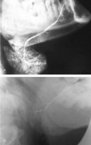
A healthy person in the picture has a homogeneous tissue structure, good visualization and distinct outlines. Also, the glands should be proportional in their size.
Indicators are considered normal if they are in the following ranges.
Parotid gland:
- width - 32-42 mm;
- length - 40-50 mm.
Mandibular:
- thickness - 11-17 mm;
- length - 28-39 mm.
Sublingual:
- length - 19-22 mm;
- width and thickness - 10-16 mm.
Any departure from the foregoing points indicates the existence of an inflammatory disease or swelling. As a rule, the sialogram shows the exact information after the first examination. If necessary, doctors can prescribe auxiliary diagnostic methods( biopsy, etc.).
Price issue
The cost of a sialogram depends on the type of study, such as the medical institution where the diagnosis is carried out, and on the average, the price of the survey ranges from 500 to 4000 rubles.