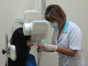
 One of the rapidly developing areas of medicine is dentistry. For the treatment and diagnosis of dental diseases, the latest technology is used. An example is the apparatus - radiovisiograph. It has some advantages, compared to the standard X-ray. Radiovisiography refers to modern methods of examination.
The study can be performed in many dental clinics available in major cities. This diagnostic procedure does not apply to expensive methods of research, it takes little time and is highly informative.
Contents
- Features of the study
- Indications and contraindications for the
- procedure Advantages of the
- procedure Technique for the implementation of the
- Evaluation of the results
Features of the study
Radiovisiography is an X-ray study used in dentistry. The device was invented more than 20 years ago, however in some clinics 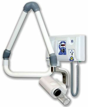 has been used since recent time.
The peculiarity of this method is that X-rays do not fall on the film, but on a special photodiode sensor. The received signal is quickly converted into an image on the computer.
With this machine, you can perform negative, positive and color images of the teeth. Depending on the indications for the study, it is possible to obtain both a target image of the tooth and a panoramic image of the oral cavity.
The filmless technology allows to reduce the time of the research. The doctor can evaluate the results of radiovisiography immediately after the procedure.
Indications and contraindications for the procedure
The method of radiovisiography is used for the diagnosis of dental diseases. Indications for the study:
- caries;
- inflammation of the tooth tissue - pulpitis;
- periodontitis;
- foreign bodies in the channel;
- neoplasm in the oral cavity.
Diagnostic procedure allows not only to make a correct diagnosis, but also to control the results of dental treatment. Thanks to the 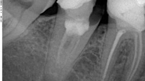 radiovisiography, it is possible to evaluate the quality of filling, implant placement.
Compared with radiology, this method is considered safer for health. Therefore, there are fewer contraindications to this method.
Dentists are inclined to believe that radiovisiography is perfectly safe for the body, so it can be performed during pregnancy( in the second trimester) and lactation. Relative contraindications include severe neurological pathologies and mental illness.
Advantages of the
method The digital diagnostic method has advantages over conventional radiography. These include:
- reduction of radiation load on the human body by 10 times;
- speed of taking pictures, so in one minute you can make several images;
- no need for a photo lab;
- the ability to store images on a computer with a doctor;
- computer image processing, if necessary, the dentist can change the brightness, contrast and color of the image;
- is convenient in evaluating the results, so the doctor has the opportunity to increase the image several times.
With the help of a radiovisiograph, it is possible to obtain panoramic shots. Due to the reduction in radiation exposure, pregnant and lactating women may be examined.
Technique for Exercise
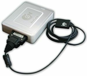
Dental Radiovisiograph RX2 HP
No special training required before conducting a study. Computer radiography can be performed at any time of the day, regardless of the intake of food and other factors.
The procedure is painless and non-invasive. The patient is asked to sit in a chair and get as close to the apparatus as possible. To protect from X-rays, put on a lead apron. The patient needs to cover the digital sensor with the mouth. To do this, he is asked to bite his teeth. Within a few seconds the patient should not move.
In most cases, the apparatus is used to obtain an image of a specific portion of the jaw in which the pathological process is localized. In some cases, dentists recommend taking a panoramic shot.
People who have undergone anesthesia may not always be able to perform the test the first time. Conducting radiovisiography is desirable after the return of sensitivity in the oral cavity.
Evaluation of results
The results of the study are ready almost immediately after the procedure. The resulting image may differ slightly from the X-ray image, since the spatial resolution of the radiovisiograph is lower. This defect is compensated by the ability to process images - adjust sharpness, contrast, etc.
Thanks to this method of diagnosis, it is possible to visualize the structure of the tooth tissue, the depth of the lesion, the presence of an inflammatory process. According to the picture, it is possible to judge what treatment intervention is required in a particular case.
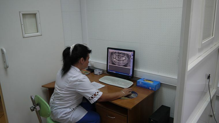
The affected tissues and neoplasms( cysts, granulomas) in the picture are of a dark color, and foreign bodies( fillings, prostheses) are represented by light areas. Normal fabric is gray.
The price of radiovisiography depends on the choice of the clinic. The price of this procedure is approximately 2 times higher than the cost of X-rays. It ranges from 150 to 400 rubles per 1 tooth.
