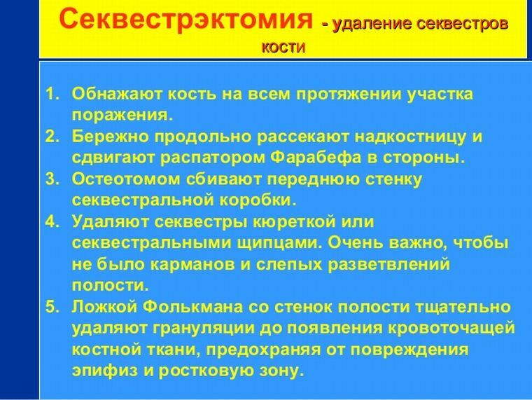
 Sequestrectomy( sequestroma) performed by a dental surgeon is a measure necessary to restore the normal functions of the dental system and the patient's health in general.
For it is aimed at the liberation of the body from an acute or chronic microbial infection, the focus of which is osteomyelitis, localized in the bowels( in the version of dental surgery - in the bones of the upper or lower jaw).
What happens if the osteomyelitis focus - bone sequestrum is not opened and cleaned? Further slow purulent fusion of the jaw bone will lead to an increase in the volume of the pathological cavity. And all this time its walls will be steadily thinning out. Until there is a jaw fracture.
All this time, poisoning of the body with toxic products of tissue decay will occur, leading to a slowdown in the work of organs and systems and a drop in the viability of the body.
Therefore, sequestrectomy should be carried out necessarily and urgently. And - radically, with scraping the bone "to a crunch," until the surgeon's instrument reaches the limits of healthy tissues, the regeneration of which will gradually fill the defect that has arisen.
When the area of necrosis is left( pass), the infection that has survived in it will continue to destroy the bone with pus from the inside, with its periodic breakthrough outwards along the fistulous passages that it has pierced.

Contents of
- Is it shown or contraindicated?
- Synthesis of technology and art
- Applied techniques and techniques
- In the rehabilitation phase of
- Complexities are possible. ..
Is it shown or contraindicated?
Excision-scraping of sequesters is indicated in the presence of a focus of both acute and chronic osteomyelitis with the formation due to its existence:
- ulcerative defects of the oral mucosa;
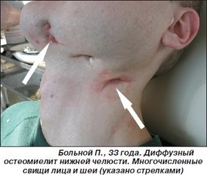
- fistulas as in the oral cavity, as on the surface of the skin of the face;
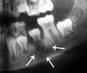
- of purulent cavities and sequesters within the jawbone;
- of the false joint due to incomplete jaw fracture( with preservation of the cortical closure plate).
Indications for immediate sequestrosis are:
- malignancy of a bone-damaged process;
- disorders of the function of organs and body systems due to chronic intoxication.
Contraindication of the intervention and with:
- the danger of exacerbation of the patient's chronic illnesses due to any general surgical operation;
- the severity of the patient's overall health, due to the presence of an exhausting disease or process.
Therefore, it is only the dental surgeon who is able to adequately assess both indications and contraindications to the operation.
Synthesis of technology and art
Surgical dentistry realizes the same principles as general surgery: 
- the greatest possible shudder of the patient's health - the operation in the shortest possible time;
- maximum thoroughness of processing the operated area, allowing to avoid the occurrence of complications;
- creation of sufficient operational access with the scope of the operational field to control the purity of processing the intervention zone.
In the case of sequestrectomy, this is:
- a thorough preliminary examination of the pathological focus by an X-ray method;
- sufficient operative access, both for the manufacture of the operation, and the subsequent drainage of the pathological cavity;
- necrectomy to completely healthy tissues while maintaining a maximum of viable bone tissue;
- , a careful treatment of the granulation tissue that appeared after the intervention:
- delayed plastic surgery of the resulting bone defect after complete elimination of the risk of reinfection.
Applied Techniques and Techniques
Depending on the dislocation of the pathological cavity, operational access is used:
- from the oral cavity of ( when osteomyelitis is affected by the alveolar process);
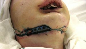
- from the extraoral section ( in the case of the vastness of the focus, with the capture of the body of the lower jaw and its branch).
After the application of anesthesia appropriate to the case( conductive local or general anesthesia), an operating access is created:
- or an incision from the oral cavity;
- or by cutting the skin parallel to the lower edge of the lower jaw( with the expectation of reaching its inner surface).
In the following steps, regardless of the access method, similar actions are performed:
- exposure of the jaw bone by detachment of the mucosa and periosteum with a single flap;
- opening of the bone cavity;
- necroctomy - scraping the cavity with a curette to healthy tissues, with the removal of sequesters and granulations, not being recovered entirely, large sequesters, fragmenting into smaller ones by cutting pliers or scissors;
- filling the cleared cavity with biologically active material, not only stimulating bone regeneration, but also having antiseptic activity;
- superposition on the mucosal-periosteal flap of deaf sutures returned to the site.

X-ray sequestration
Cavity drainage is performed by solutions of antiseptics through a catheter left in a postoperative wound, the wound itself is also treated with antiseptics: a solution of brilliant green or Chlorhexidine to prevent suppuration and is isolated by the application of an antiseptic dressing. The lower jaw is usually immobilized with a plaster or plastic detachable lint.
Seams are removed after 6-8 days after the operation.
When an osteomyelitis of the upper jaw occurs with the involvement of the maxillary sinus at the same time with the sequestrectomy of the jaw, a radical healing of the maxillary sinus is performed.
At the stage of rehabilitation
After the completion of the operation phase, steps are taken to:

After the operation, the appearance, as well as the feeling is not very. ..
- prevention of infection:
- stimulation of bone regeneration by the appointment of calcium-containing agents;
- general strengthening of the patient's health and raising the protective capabilities of his body.
Antibacterial and desensitizing therapy, with the use of analgesics if necessary, as well as the use of physiotherapeutic( ultrasound therapy, iontophoresis with zinc and copper) and similar therapies( laser therapy), vitaminization of the body serve these purposes.
Complexities are possible. ..
On the way to full recovery, the following problems may become:
- trabecular-spongy structure of the jawbone with a lot of micro-holes and micro-pores, an abundance of blood vessels giving constant bleeding during the operation, which does not allow to achieve the proper level of visual control;
- long duration of existence of the focus of chronic osteomyelitis, leading to irreversible changes in bone structure;
- is an exhausting disease.
For these and other reasons, the production of an operation with a sufficient level of radicality is not always possible, and the surviving microfouca of infection is always ready to cause a relapse of the development of purulent-putrefactive osteomyelitis in the bowels of the jaw that leads to its fracture, malignancy, and chronic intoxication of the patient's body.
