The eye apparatus is stereoscopic and in the body is responsible for the correct perception of information, the accuracy of its processing and further transfer to the brain.
The right part of the retina, through transmission through the optic nerve, sends to the brain information of the right part of the image, the left part transmits the left share, eventually, the brain connects both, and a general visual picture is obtained.
This is binocular vision. All parts of the eye form a complex system that performs an action for the qualitative perception, processing and transmission of visual information in electromagnetic radiation.
- 1. External structure of the human eye
- 2. Internal structure of the
- 3. Principle of operation of the human eye
- 4. Diseases
- 5. Prevention
External structure of the human eye
The eye consists of the following external parts:
- Eyelids.
- The lacrimal department.
- Eyeball.
- The pupil.
- Cornea.
- Sclera.
Eyelids
Protect the eyes from the negative effects of the environment. They also protect against accidental injuries. The eyelids consist of muscle tissue, which is covered from the outside with the skin, and inside they are covered with conjunctiva, in the form of a mucous membrane. Muscle tissue provides free, moistened motion to the eyelids.
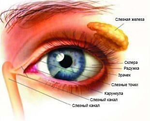
The conjunctiva has a moisturizing effect, which causes a smooth glide of the eyelid along the eyeball. At the edge of the eyelids, eyelashes are arranged, which also perform a protective function for the eye.
The lacrimal section of the
Includes the lacrimal gland, additional glands and pathways that serve as an outlet for tears. The lacrimal gland is located in the pit outside the eye socket in the upper corner.
The lacrimal pathways are located on the inside of the corners of the eyelids. Additional glands are formed in the arch of the conjunctiva, as well as near the upper edge of the cartilage of the eyelid.
Tears from the accessory glands serve as a moisturizing substance for the cornea and conjunctiva. They purify the conjunctival sac of foreign bodies and microbes.
Approximate amount of excreted tears per day is 0.4-1 ml. When irritation of the conjunctiva begins to work tear gland. Blood supply of the gland gives a tear duct.
Pupil
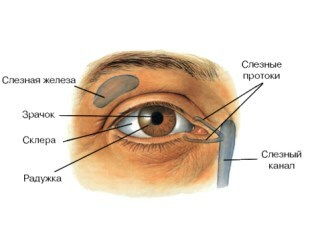
Located in the center of the iris of the eye and is a round hole with a size of 2 mm and up to 8 mm. The visual energy generated in the mesh shell is formed by passing through the pupil into the eye of the light rays.
The pupil has the property of expanding and narrowing, depending on the influence of illumination. Luminous flux falls on the retina of the eye, and it transfers this information to the nerve centers that optimally regulate the pupil's work.
This function is provided by the muscles of the iris - the sphincter and dilator. The sphincter serves to narrow the pupil, dilator for enlargement. Due to this property of the pupil, the visual function of the eye does not suffer from bright sun or fog.
The change in the diameter of the pupil is automatic and completely independent of personal desire. In addition to a bright light flux, a decrease in the pupil can cause irritation of the trigeminal nerve and drugs. The increase causes strong emotions.
Cornea
The cornea of the eye is an elastic membrane. It is of transparent color and is a fraction of the refractive apparatus, consists of several layers:
- epithelial;
- Bowman's membrane;
- stroma;
- descemet membrane;
- endothelium.
The epithelial layer protects the eye, normalizes the hydration of the eye and provides it with oxygen.
The Bowman membrane is located under the epithelial layer, its function in providing eye protection and nutrition. The Bowman membrane is the most non-renewable.
Stroma is the major part of the cornea that contains collagen horizontal fibers.
Read more - the price of Zovirax ointment. How much does a drug in the CIS cost?
In the news( here) reviews about Timolol.
Description of eye drops Okumil!http: //moezrenie.com/lechenie/ kapli-dlya-glaz / okumetil-opisanie.html
The descemet membrane serves as the separating substance of the stroma from the endothelium. It is very elastic, which is rarely damaged.
Endothelium in the cornea serves as a pump for the outflow of excess fluid, as a result of which the cornea remains transparent. Endothelium also helps in nourishing the cornea.
It is poorly restored, and the number of cells filling it decreases with age, and along with them the transparency of the cornea decreases. The density of endothelial cells can be affected by injuries, illnesses and other factors.
Give a breather to your eyes - see the video on the subject of the article:
Sclera
Is the outer shell of the eye, which is opaque .It smoothly passes into the cornea. The oculomotor muscles are attached to the sclera, and it itself contains vessels and nerve endings.
Internal structure
Let's analyze, the inner structure of the eye:
- Lens.
- Vitreous humor.
- Cameras with watery moisture.
- Iris.
- Retina.
- The optic nerve.
- Arteries, veins.
Crystal
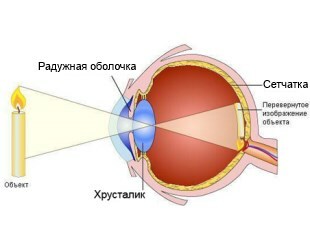
It has an accommodation mechanism, and it is similar to a biological lens, which has a biconvex shape. The lens is behind the iris, behind the pupil and has a diameter of 3.5-5 mm. The substance from which the lens consists consists of a capsule.
Under the top of the capsule is a protective epithelium. In the epithelium there is a property of cell division, due to the densification of which with age, hyperopia appears.
The lens is fixed with thin threads, one end of which is tightly woven into the lens, its capsule, and the other end is connected to the ciliary body.
When the tension of the threads changes, the accommodation process takes place. The lens is devoid of lymphatic vessels and blood vessels, as well as nerves.
It provides the eye with light and refraction, gives it the accommodation function, and is the eye divider to the back and front sections.
Vitreous Body
The vitreous humor of the eye is the largest formation. This substance without the color of the gel-like substance, which is formed in the form of a spherical form, is flattened in the sagittal direction.
The vitreous humor consists of a substance of a gel-like substance of organic origin, a membrane and a vitreous channel.
Before him is the lens, the zonular ligament and the ciliary processes, the posterior part of it closely approaches the retina. The connection of the vitreous and retina occurs in the optic nerve and in the part of the dentate line where the flat part of the ciliary body is located. This area is the base of the vitreous body, and the width of this belt is 2-2.5 mm.
The chemical composition of the vitreous: 98.8 hydrophilic gel, 1.12% dry residue. When a hemorrhage occurs, the thromboplasty activity of the vitreous humor increases dramatically.
This feature is aimed at stopping bleeding. In the normal state of the vitreous body, there is no fibrinolytic activity.
Nutrition and maintenance of the vitreous environment is provided by diffusion of nutrients, which through the vitreous membrane, enter the body from the intraocular fluid and osmosis.

Note - eye drops of Travatan. Review of the drug, its prices and analogues.
In the article( link) instructions for use on eye drops Taurine.
The price of Indocollar!http: //moezrenie.com/lechenie/ kapli-dlya-glaz / indokollir-bystraya-pomoshch-pri-bolyakh-glaz.html
There are no vessels and nerves in the vitreous humor, and its biomicroscopic structure represents various forms of gray ribbons with white specks. Between the tapes are areas without color, completely transparent.
Vacuoles and opacities in the vitreous body appear with age. In the case where there is a partial loss of the vitreous humor, the space is filled with intraocular fluid.
Cameras with watery moisture
The eye has two chambers that are filled with watery moisture. Moisture is formed from the blood by the processes of the ciliary body. Its allocation occurs first in the anterior chamber, then it enters the anterior chamber.
In the front chamber watery moisture enters through the pupil. In a day the human eye produces from 3 to 9 ml of moisture. In watery moisture, there are substances that nourish the lens, endothelium of the cornea, the anterior part of the vitreous body, and also the trabecular network.
It contains immunoglobulins that help remove harmful factors from the eye, its internal part. If the outflow of watery moisture is disturbed, then this can develop an eye disease, like glaucoma, and also increase the pressure inside the eye.
In cases of impaired integrity of the eyeball, the loss of aqueous humor leads to hypotension of the eye.
Iris
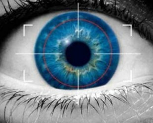
Iris is the avant-garde department of the vascular tract .It is located immediately behind the cornea, between the chambers and in front of the lens. The iris is round and located around the pupil.
It consists of a boundary layer, a stromal layer and a pigment-muscle layer. It has an uneven surface with a pattern. In the iris, pigment cells are present, which are responsible for the color of the eyes.
The main tasks of the iris: the regulation of the light flux, which passes to the retina through the pupil and the protection of photosensitive cells. From the correct functioning of the iris depends visual acuity.
The iris has two muscle groups. One group of muscles is located around the pupil and regulates its decrease, the other group is located radially along the thickness of the iris, regulating the dilatation of the pupil. Iris has many blood vessels.
The
retina is the optimal thin layer of nerve tissue and represents the peripheral part of the visual analyzer. In the retina there are photoreceptor cells that are responsible for perception, as well as for the transformation into nerve impulses of electromagnetic radiation. It adjoins from the inside to the vitreous body, and to the vascular layer of the eyeball - from the outside.
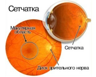
The retina has two parts. One part is visual, the other is the blind part, which does not contain photosensitive cells. The internal structure of the retina is divided into 10 layers.
The main task of the retina is to receive the light flux, process it, translate it into a signal that forms a complete and coded information about the visual picture.
Optic nerve
The optic nerve is an interlacing of nerve fibers. Among these thin fibers is the central channel of the retina. The initial point of the optic nerve is in the ganglion cells, then its formation occurs through passage through the scleral membrane and fouling of nerve fibers by meningeal structures.
The optic nerve has three layers - solid, arachnoid, soft. Between the layers is a liquid. The diameter of the optic disc is about 2 mm.
Topographic structure of the optic nerve:
- intraocular;
- is intra-orbital;
- intracranial;
- intracanular;
The principle of the human eye
The light stream passes through the pupil and through the lens is focused on the retina. The retina is rich in photosensitive sticks and cones, which in the human eye are more than 100 million.
Video: "The vision process"
Wands provide sensitivity to light, and cones give the eyes the ability to distinguish colors and small details. After refraction of the light flux, the retina transforms the image into nerve impulses. Further, these impulses pass to the brain, which processes the information that has arrived.
Diseases
Diseases associated with a violation of the structure of the eyes can be caused both by an incorrect arrangement of its parts relative to each other, and by internal defects of these parts.
The first group includes diseases that reduce visual acuity:
- Myopia. It is characterized by an enlarged eyeball in comparison with the norm. This leads to the focusing of light passing through the lens, not on the retina, but in front of it. The ability to see objects that are far from the eyes is impaired. Nearsightedness corresponds to a negative number of diopters in measuring visual acuity.
- Farsightedness. It is a consequence of a decrease in the length of the eyeball or loss of elasticity of the lens. In both cases, the accommodative possibilities are reduced, the correct focusing of the image is violated, the light rays converge behind the retina. Violated ability to see objects located near. Farsightedness corresponds to a positive number of diopters.
- Astigmatism. This disease is characterized by a violation of the sphericity of the eye shell due to defects in the lens or cornea. This leads to an uneven convergence of the rays of light entering the eye, the clarity of the image obtained by the brain is violated. Astigmatism is often accompanied by myopia or hyperopia.
Pathologies associated with functional impairment of certain parts of the organ of vision:
- Cataract. With this disease, the lens of the eye becomes cloudy, its transparency and ability to conduct light are violated. Depending on the degree of turbidity, visual impairment may be different up to complete blindness. Most people have cataracts in old age, but do not progress to severe stages.
- Glaucoma is a pathological change in intraocular pressure. It can be provoked by a variety of factors, for example, a reduction in the anterior chamber of the eye or the development of cataracts.
- Myodesopsy or "flying flies" before the eyes. Characterized by the appearance of black dots in the field of view, which can be represented in different numbers and sizes. Points arise because of disorders in the structure of the vitreous. But this ailment causes not always physiological - "flies" can appear due to overwork or after the transfer of infectious diseases.
- Strabismus. It is provoked by a change in the correct position of the eyeball in relation to the eye muscle or a violation of the eye muscles.
- Retinal detachment. The mesh and the posterior vascular wall are separated from each other. This is due to a breach of the tightness of the retina, which occurs when tearing its tissues. The detachment is manifested by the clouding of the outlines of objects before the eyes, the appearance of flares in the form of sparks. If individual angles fall out of the field of view, this means that the detachment has taken heavy forms. In the absence of treatment, complete blindness occurs.
- Anophthalmus - insufficient development of the eyeball. A rare congenital pathology, the cause of which is the violation of the formation of the frontal lobes of the brain. Anaphthalmus can also be acquired, then it develops after surgical operations( for example, to remove tumors) or severe eye injuries.
Prevention
To keep the vision clear for many years will help the following recommendations:
- It is necessary to take care of the health of the circulatory system, especially the part that is responsible for the flow of blood to the head. Many visual defects arise from atrophy and damage to the eye and brain nerves.
- Eye contact must not be obstructed. When working with the constant consideration of small objects, you need to make regular breaks with the conduct of eye gymnastics. The workplace should be arranged so that the brightness of the lighting and the distance between objects are optimal.
- The intake of enough minerals and vitamins into the body is another condition for maintaining healthy eyesight. Especially for the eyes, vitamins C, E, A and minerals such as zinc are important.
- Proper eye hygiene can prevent the development of inflammatory processes, complications of which can significantly impair vision.
