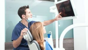
 The diagnosis and treatment of teeth in the observation of serious diseases is carried out using X-rays. Diagnosis is used to obtain more accurate data on possible pathological processes.
Without X-ray examination, the dentist needed to open the tooth and gum to understand what caused the abnormal condition.
X-ray of the teeth allows you to make a complete clinical picture and see the anatomical features of the structure of the patient's jaw. This is very important for effective and full-fledged treatment.
X-ray examination of teeth is performed in all medical institutions. No serious procedure for the treatment or removal of teeth can not be carried out without such a diagnosis.
The study can be performed to obtain a picture of both a single tooth and a certain part of the jaw. The picture also shows a soft gum tissue that can be affected by inflammation.
Contents of
- Indications for procedure
- Diagnosis without excess blood
- X-ray during pregnancy
- Small patients - special approach
- How often can radiography be done?
- Additional recommendations
- study options
- preparation
- X-ray preparation
- survey description How harmful is the
- procedure What patients think
- Question price
Indications for
procedure An external examination performed by a dentist at every patient's reception does not always allow to establish precisely the reason for the occurrence ofa pathological condition. To properly determine the diagnosis and the method of treatment, use an x-ray machine.
Indications for the procedure are the following deviations from the norm of the teeth: 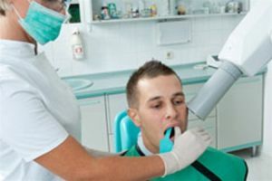
- abnormal position of the dentition;
- a latent cavity formed as a result of caries;
- periodontal disease;
- pathological processes occurring under seals or crowns;
- injury to internal tissues of the tooth or jaw;
- presence of neoplasms or abscesses;
- implant installation.
Diagnostic results facilitate the work of a specialist, giving him the opportunity to accurately determine which method to apply therapy, or to resort to tooth removal. X-rays can be given in the presence of other diseases to determine their course.
Diagnosis without excess blood
Most procedures that involve penetrating the inside of the tooth and gums can not be carried out without preliminary x-ray diagnostics.
The image is taken to determine the condition of bone tissue, roots, as well as the presence of caries under the crown( seal) or in the spaces between the teeth. The device is able to show the state of soft tissues inside the gums, to identify possible inflammation and cracks in the canals.
Radiography allows you to accurately determine the place where it is necessary to carry out manipulations to eliminate pathology. The doctor will not need to do unnecessary actions, which can bring painful feelings to the patient or lead to complications.
X-ray examination - the possibility of establishing the right action plan for a specialist to treat the disease.
X-ray during pregnancy
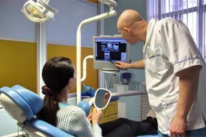 Dentists should not prescribe a diagnosis to women in the first trimester of pregnancy. After this period is over, the x-ray of the teeth is done only in cases of acute necessity, when it is impossible to treat without it.
To reduce the radiation load, specialists need to use a special film( E-class).It is recommended to use a digital method that will not cause any harm to the woman and her fetus.
It is acceptable to have a tooth X-ray and during breast-feeding. Since the dose of radiation is small, breast milk does not accumulate any radiation, and accordingly the child's body will not suffer.
In the first trimester, such a diagnosis is contraindicated.
Small patients - a special approach
X-ray images of infant teeth are very rare, only with serious pathological processes that occur inside the gums or tooth. The procedure provides an opportunity to get rid of violations that will affect the formation of permanent dental makeup.
Diagnose with the application of a minimum dose of radiation. Before the procedure begins, the child is protected, using a special apron made with particles of lead. Reduce the negative impact of the device can be, if carried out a digital study.
How often can I take a radiograph?
The frequency of X-ray diagnostic procedures is determined by the position of SANPIN( 2.6.1.1192-03).This provision determines the maximum dose of radiation for prophylactic purposes and treatment. How often you can do the survey depends on the equipment that is being used.
The most safe method is a digital study of the state of dental tissues. As little as possible should be X-ray on a film device.
Any procedure has a negative effect on the body, therefore it is recommended to do radiography only when necessary.
Additional recommendations of
X-rays cause harm to the body, albeit insignificant. You can reduce the risk if you choose a good clinic that is equipped with modern equipment.
Do not give up X-ray of baby teeth in children or during pregnancy. If the dentist assigns such a diagnosis, then it is necessary.
Varieties of research
In recent years, the radiography of dental tissues has been increasingly used. This is due to the development of devices that allow you to receive instant and accurate pictures. Thus, treatment is faster, and patients experience minimal discomfort.
Diagnosis can be performed using old and new techniques. Depending on the equipment used, there are four types of X-ray of the teeth:
- bite: to identify caries and calculus;
- aiming: to determine the internal state of the tooth and gums;
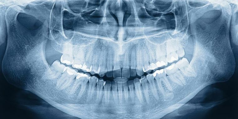
- panoramic: to get a more accurate picture of the overall condition of the jawbone;
- digital: to obtain a clear image of both a single tooth and the entire dentition.
The latest type of tooth diagnosis is 3D X-ray. This method of research allows you to get a panoramic or three-dimensional snapshot, 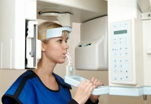 which is displayed on the computer screen.
As a result of image processing, the doctor receives the most accurate picture.
In order not to have to go through the diagnostics again and the picture turned out to be of high quality, the examination should be carried out according to certain rules, which must be observed not only by the medical specialist, but also by the patient himself.
Preparation for X-ray
Before starting the procedure, the patient must remove all ornaments that are on the face, head or neck.
Metal objects can distort images or appear as a "shadow".As a result, the dentist can be confused, and the patient will need to undergo a second diagnosis.
Survey description
The radiography procedure is performed according to a certain scheme, however, in some medical institutions, depending on the apparatus used, the survey process may differ.
So, as usual, do the X-ray of a tooth:
- the patient's body is covered with a special apron;
- the patient goes inside a special apparatus;
- bites the plastic wand;
- lips closed;The
- is pressed against the platform by the chest.
The position of the person should be level. In some cases, the head needs to be rotated to get an image of a particular section. After the position of the body is taken, take a picture.
How harmful is the procedure
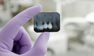 Any radiation is harmful to the body. But the development of diseases occurs only with a large dose of radiation.
X-ray of the tooth has an effect on a person in such small doses that it can not provoke pathological processes.
If the patient has any doubts, it is necessary to consult a doctor. However, it should be understood how important such a diagnosis is, and what health problems may arise when refusing it.
What patients think
From the practice of patients in dental clinics.
I went to the dentist with complaints about pain in the gums. To determine the cause, it took an X-ray. As a result, it turned out that a cyst was formed inside the gums. I had to remove the tooth. At the same time in another clinic, where the dentist conducted an examination, they offered to treat.
Maria
I'm in the fifth month of pregnancy. One of these days the tooth was strongly ill. I went to the doctor. They offered X-rays. At first she was frightened, but the doctor explained that it was safe. It turned out that the cause was a carious cavity inside the tooth.
Natalia
My dentist( four years old) prescribed a tooth X-ray. Initially I wanted to refuse, but then I consulted with a specialist and decided that the damage from x-rays is much less than from the lack of proper treatment.
Tatiana
The price of the question
The price of the X-ray of the tooth depends on several factors. The main role is played by the apparatus, which is used for diagnostics. 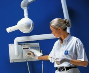
Also, the cost may depend on the type of picture, the area of examination, and, of course, from the medical institution itself. On average, the cost of radiographing the tooth is from 250 to 1500 rubles.
X-ray - examination of teeth is an important stage in the diagnosis of dental diseases. It is not always possible to conduct quality treatment without special tissue testing.
With the help of an X-ray, you can correctly diagnose for orthodontic, surgical and therapeutic procedures.
