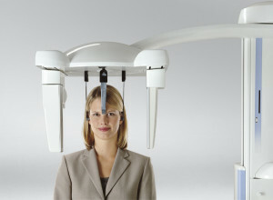
 Teleradiography( TRH) is a kind of radiography, but it differs in that it is done from a remote distance. Therefore, you can get a fairly clear and accurate image, corresponding to the real size of the area under investigation.
Most often this type of diagnosis is used in dentistry. The teleradiogram is the main assistant for orthodontists and is assigned to all patients, before the installation of braces or other orthodontic structures. TG is the most important procedure before the beginning of orthodontic treatment.
After all, the most effective therapy plan can be made up only by revealing all the features of the problem area: the ratio of the sizes of bones and soft tissues, the exact location of the teeth, the angles of inclination and the degree of their displacement. Also, a head shot is taken during correction, or after its termination( to control changes).
Teleradiography for dentists( and especially for orthodontists) is the most informative type of diagnostics, therefore it has wide application.
Contents
- Usage in dentistry
- Indications and contraindications
- Kinds of telecasts
- How diagnostics are carried out
- Advantages of
- method Viewpoint
- Cost of service
Use in dentistry
TGG is assigned to a patient with the purpose of:
- to determine the size of cranial bones, their structure, location, connection;
- to detect a focus with abnormal tissue growth, its location;
- determine the direction of tooth dislocation;
- find out the location of the tissues in relation to the skull;
- to find the factors on which the wrong bite depends( the structure of other bones of the skull);
- identify abnormalities that are difficult to treat;
- find out the features of the anatomical structure;
- track the dynamics of growth;
- be convinced of the effectiveness of the treatment.
Indications and contraindications
Indications for use of this type of diagnosis for children and adult patients are the same. 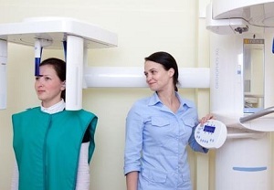
Indications:
- the most widely used procedure of TGH was found in orthodontics with correction of occlusion;
- the statement of the correct diagnosis, or its specification;
- planning and prognosis of treatment of various dentoalveolar anomalies;
- monitoring of the ongoing processes of changes in the tissues of periodontal disease in dynamics and control over the stabilization of the results achieved.
Radiation load for this test is minimal and is considered harmless for personnel and patients, but nevertheless it is not recommended for pregnant women of TRH.
In addition, this study has several more contraindications:
- bleeding and / or pneumothorax;
- the patient's general severe condition;
- pregnancy( especially 1 and 3 trimester).
With the help of TRG, you can make a fairly clear panoramic picture of the skull in profile and facet. Separate 3 types of such diagnostics.
Kinds of the telecast of the
Specialist, depending on the goals, one or several pictures are needed for a full-fledged diagnostics:
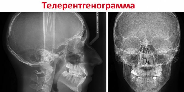
- Frontal .This type of research is used for diagnostic purposes before starting orthopedic treatment and is the main one. A picture of the head is taken from behind and in front. It allows you to see quite clearly the existing inflammatory processes, facial asymmetry and fractures.
- TRG in the lateral projection is required for patients with malocclusion. With the help of a lateral snapshot it is possible to reveal the pathology of the dentoalveolar system structure as accurately as possible and make an optimal plan for correction. Research in this projection allows you to determine the angles of inclination of the lower anterior teeth, the peculiarities of their location, and also to control the treatment and calculate its duration. Anyone who plans to correct the bracket system, of course, necessarily make such a picture.
- Axial ( or chin) teleradiography is assigned only as an additional diagnostic in conjunction with other TWG species. Such a study gives the most complete picture of the shape and structure of the head in three dimensions. This is necessary to determine changes in the structure of the nasal cavity, maxillary sinus and cheekbones. It is used, as a rule, when implantation of the upper anterior teeth is required.
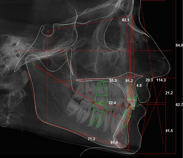
Side image of the head, obtained with the help of the TRG
Regardless of the projection in which the image is required, the diagnostic procedure is quite simple.
How is the
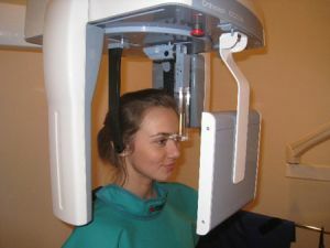
diagnosed? Usually, the shooting is performed while standing, but if the patient can not stand for some reason, a sitting position is allowed.
During scanning it is very important to take and fix the correct, natural position of the head( for clarity of the image).In order to avoid movement, special plastic stops are inserted into the ear passages.
Before the procedure, the patient's face on the middle soft tissue line( including the soft palate) is smeared with barium suspension( to achieve contrast).
The procedure does not cause unpleasant sensations in the subject and takes only a few minutes. The doctor should carefully analyze the finished image and carry out its decoding.
Teleradiography is of key importance in modern orthodontics. Without TGF, the diagnosis and correction of bite pathologies is not possible. Wide application in orthodontics this procedure of diagnostics has received due to many advantages.
Decoding with the help of a special computer program:
Advantages of the
 technique The head image is as close as possible to the real dimensions, which allows to get a full understanding of this or that dentoalveolar pathology.
Also the study is quite affordable, and the result will be ready in a few minutes.
The procedure is safe for patients, because modern medical equipment has a minimum dose of radiation.
In comparison with other types of diagnostics, the picture obtained with the help of TRH has practically no drawbacks. But it is worth considering that the accuracy of the result depends not only on the apparatus, but also on the literacy of the specialist who deciphers the results.
Viewpoint
Those patients who did the teleradiogram generally leave positive feedback.
The problem of crooked teeth has haunted me since childhood. He was always shy to smile, and to go to doctors was afraid. But at the age of 21 I decided to correct the occlusion. I went to an orthodontist in a private clinic, he immediately sent me to take a picture in two projections( front and side TRG).I'm surprised at how far forward medicine has gone.
For some 7 minutes I got a clear snapshot of all the teeth from different angles. In addition, they say that teleradiography is safe for health.
Kirill, 36
My son( 8 years old) with an anomaly of bite orthodontist first of all appointed TRG.I was worried about the fact that the child will be doing x-rays, because it's not safe.
But the doctor convinced me that this study differs from the usual X-ray, the radiation load is minimal and absolutely harmless.
I also feared that the result might turn out to be distorted, inaccurate, since the child is unconcerned. But, as it turned out, in vain.
The procedure was rather fast, no more than 8-10 minutes, the son was able to stand this time motionless, especially since the head was fixed. Everything was painless and noiseless. We got the picture almost immediately. And there was such a diagnosis, contrary to expectations, inexpensive.
Alina, 35
Cost of service
The price range in different clinics for the service of teleradiography varies from 800 to 3900 rubles.
This difference is due to several factors: the type of diagnosis, qualification and experience of the specialist, the equipment and quality of equipment, the type of subscription and the discount system for regular customers, as well as the prestige and location of the clinic.
As a rule, the price of research includes recording the image on the disc and printing it.
