 In order to have exhaustive information about the condition of the oral cavity, it is not enough for the dentist to see only the tip of the dental "forest", limited to the fact that the teeth are from the outside - he needs a complete picture of the dental horizon.
And not only that rises above the gums, but also the "underground" hidden in the depths of these jaw joints. Therefore, dentists widely use the method of radiography - a technique that fixes and demonstrates on the X-ray image the complete structure of this hard and impermeable tissue.
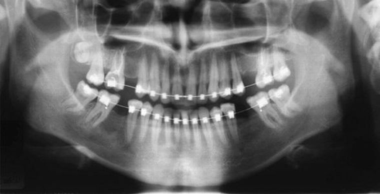
A circular x-ray taken for this purpose with the capture of both dental arches( both upper and lower) is called a panoramic one.
It shows a picture unfolded on the plane, demonstrating - in the entire width of their rows - not only the state of the teeth outside, but also their "thickness" - deep structure, internal structure, and the state of their roots.
Contents of the
- What is the purpose of the
- study? How is the
- panorama survey done?
- quality price. Arguments and conclusions.
. What is the purpose of the
research? A panoramic image allows you to find out the exact number of teeth. After all, not all of them come to the surface - there are rudiments-bookmarks, there are wisdom teeth, "sleeping" until their term in the thickness of the jaw, there are teeth that must be cut, but for some unknown reason they do not erupt.
Which one? Will they break through at all? When? All these questions can be answered by examining a panoramic snapshot of the jaw.
In addition to the quantity, the dentist uses a panoramic snapshot to evaluate the relative position of the teeth:
- In some cases, two of them "claim" one hole of the , interfering with each other both in development and in operation.
- Either they are located too far apart from each other , forming significant free spaces. How to distribute them more evenly, what method to apply for this? Will the use of a kapa be sufficient or will serious surgical intervention be required?

Panoramic tooth image with braces
Also a panoramic X-ray of teeth is done if:
- The question of removal( extraction) of the tooth is raised. Will it be sufficient for this purpose to use forceps or will it be necessary to cut the gums so that its roots are easily removed without fragmentation? What is the structure of the roots of neighboring teeth? Will they prevent the removal process? Acute toothache .Outside, the tooth looks healthy, it is sealed, its internal contents are reliably isolated from external influences. But this does not mean that the infection inside it did not penetrate from the other side - with the blood flow through the vascular-neural bundle entering the apex of the root, into which the artery also enters. And the tooth, remaining untouched from the outside, is struck from the inside. Or maybe the pain causes a tumor growing in the thickness of the jaw? What is its structure? Is it benign or malignant? How fast does it grow?
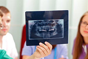
All these questions are answered by a panoramic shot. Allows him to answer the question of what the general state of each individual tooth. If a pathology is detected, other, targeted images are taken, but for a general review, evaluation of the general condition, a panorama of all the teeth present in the patient is needed at the moment.
 Panoramic X-ray provides an irreplaceable service for teeth prosthetics, as well as for large-scale maxillofacial interventions, when the whole architectonics of the facial part of the skull is completely changed( as a result, say, of a gunshot injury with destruction of the upper, lower or both jaws).
In addition to the teeth, the orthopantogram( the second name of the panoramic image) contains both temporomandibular joints in its field, as well as the nasal cavity combined with the adnexal( maxillary) sinuses( there are cases when the roots of the teeth "sprout" into it, provoking a stubborn, nottreatable, sinusitis).
Orthopantogram( as a special case of X-ray dental examination) provides:
- the possibility of diagnosing for dental problems;
- monitoring the accuracy of individual manipulations during treatment;
- the possibility of monitoring at the end of the work done.
And yet this is an opportunity to research teeth that do not hurt, but are already being destroyed or regenerated.
How is the
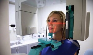
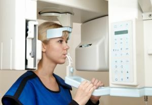
panoramic shooting performed? The procedure is that the X-ray unit "travels" along the arc at the required focal length, so that the panorama turns into a flat image on an X-ray film or it is created on a computer sensor.
The procedure does not require special preparation, is absolutely insensible to the patient and carries a minimum of radiation load.
Price quality
The price of orthopantograms in one of the dental clinics in Moscow is an average of 1300 rubles. The study is carried out on the German orthopantomograph Orthophos, equipped with a tomograph Samsung Rayscan Symphony Alpha Pano 3D South Korean production.
The equipment allows to study and record the results digitally, and this enables:
- to reduce( up to 90%) the radiation load on the organism by decreasing the duration of the irradiation time;

- acceleration of the image acquisition process and reduction of the waiting time for the result of the study( there is no need for X-ray film and re-examination when the film or reagents are matched);
- to create a 3D image of the area under investigation and its parts with an increase to the dimensions required for accurate analysis;
- easy storage, copying and instant image transmission anywhere in the world if you need to consult with foreign experts.
Arguments and conclusions of
In addition to the diagnostic and correction capabilities in the treatment process, the speed and accuracy of its implementation, there is one more reason to make an orthopantomogram: one dental patient who addresses a dental care will be much cheaper than several standard X-ray sighting shots.
And the latest argument in favor of panoramic shooting: the doctor as a specialist can see what type of research should be offered to the patient.
