
Content
- The role of the ovum in the body of a pregnant woman
- What does the fertilized egg consist of?
- How and why the fertilized egg is measured
- Size chart of the ovum in the first trimester of pregnancy
- Fetal size chart in the first trimester of normal pregnancy
- Pathology of the ovum
- Too large fertilized egg
- Fertile egg too small
- Wrong position
- Fertile egg of irregular shape
- Bubble drift
- Empty fetal egg
- Symptoms of an empty ovum
- HCG for anembryonia
- Embryonic treatment
- Complications
- Pregnancy after anembryonia
- Why the fertilized egg is not visible
- What to do if the fertilized egg does not meet the standards
The role of the ovum in the body of a pregnant woman
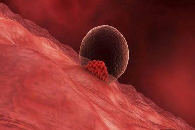
A fertilized egg is a structure that forms after the fusion of an egg and a sperm cell, transforming into a fetus and amniotic membranes. First, 2 cells are formed, then 4, 8, etc. They are transformed into a zygote, then into a blastocyst, an embryo. On the 7-10th day, the formed ovum is introduced into the uterine cavity.

The fertilized egg performs the following functions:
- preserves the developing fetus;
- provides the embryo with the necessary nutrition;
- supports blood circulation of the growing embryo at an early stage of development;
- fixes the future fetus in the uterine cavity;
- forms amniotic membranes;
- creates the basis for the formation of the placenta;
- activates the production of the pregnancy hormone - hCG.
What does the fertilized egg consist of?
The fertilized egg is oval in shape and consists of:
- external protective shell - chorion;
- internal nutrient medium - amnion;
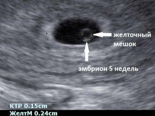
- the yolk sac, which provides embryonic nutrition at the initial stage of intrauterine development;
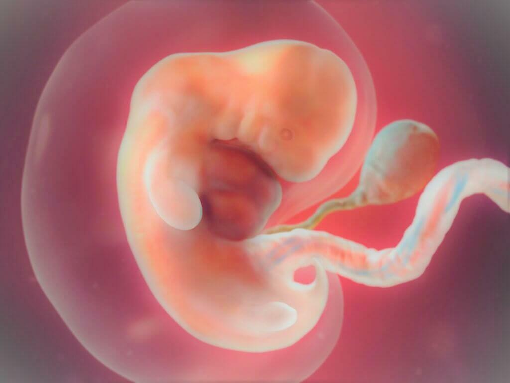
- the embryo, which is visually determined only 5 weeks after the onset of pregnancy.
It is at the very moment when a modern specialist doctor sees a fetal egg on the screen of an ultrasound machine, future mothers and fathers hear the cherished words: "You will have a child."
Knowing the normal size of the ovum by weeks of pregnancy (tables can be compiled in accordance with obstetric or actual terms) and the parameters of the fetal egg of the expectant mother in each case, it is possible to predict whether it develops correctly pregnancy.
How and why the fertilized egg is measured
The size of the ovum in the early stages of pregnancy is the main parameter by which the doctor can judge how the baby is developing. The embryo is still very small, it is not possible to measure it and its individual parts, but the growth rate of the ovum is a very informative indicator of the development of pregnancy as a whole.
The size of the ovum speaks not only of development, but also of compliance with certain obstetric terms. The fact is that at the very beginning of pregnancy, when the embryo is still emerging, there is not much difference in height and weight. It is much later that children in the womb begin to grow in different ways, in accordance with their genetic program (some are tall, others are small). In the meantime, all babies develop almost identically, so the growth rate of the ovum is almost the same.
The uncertainties and range of values in the diagnostic tables are related to the likelihood of late implantation as well as other factors that may affect the size of the ovum, but do not pose a threat to the development of the baby.
A special technique is used for measurement. An ultrasound diagnostician so lays a straight visual line through the ovum, which he sees on the monitor, so that the ends of the segment are located on opposite points of the inner shell of the fetal bag. This size is called SVD - average inner diameter.
This size is determined by the very first. Then the coccygeal-parietal size of the embryo itself is added to it. The size of the yolk sac is also considered important.
It is very bad if it is not visualized at all. If it is visible and its dimensions correspond to the norms, this still does not guarantee that the baby will be healthy, that the pregnancy will proceed without problems.
Growth rates can be seen in the table below.
Size chart of the ovum in the first trimester of pregnancy
| Deadline for the last menstruation (weeks) |
Conception period (weeks) | Interior diameter (mm) |
Square (mm2) |
Volume (mm3) |
| 5 | 3 | 18 | 245 | 2187 |
| 6 | 4 | 22 | 363 | 3993 |
| 7 | 5 | 24 | 432 | 6912 |
| 8 | 6 | 30 | 675 | 13490 |
| 9 | 7 | 33 | 972 | 16380 |
| 10 | 8 | 39 | 1210 | 31870 |
| 11 | 9 | 47 | 1728 | 55290 |
| 12 | 10 | 56 | 2350 | 87808 |
| 13 | 11 | 65 | 3072 | 131070 |
Fetal size chart in the first trimester of normal pregnancy
| The term for the last menstruation (weeks) | Conception period (weeks) | Coccyx-parietal size (mm) | Biparietal dimension (mm) | Yolk sac diameter (mm3) |
| 5 | 3 | 3 | – | – |
| 6 | 4 | 6 | – | 3 |
| 7 | 5 | 10 | – | 4 |
| 8 | 6 | 16 | 6,0 | 4,5 |
| 9 | 7 | 23 | 8,5 | 5,0 |
| 10 | 8 | 31 | 11,0 | 5,1 |
| 11 | 9 | 41 | 15,0 | 5,5 |
| 12 | 10 | 53 | 20,0 | 6,0 |
| 13 | 11 | 66 | 24,0 | 5,8 |
Pathology of the ovum
Let's look at what the pathologies of the ovum are and what the prognosis is.
Too large fertilized egg
Too large a fertilized egg in the early stages can talk about various pathologies of both the fetus itself and this pregnancy. Often, an excess of size is a harbinger of a frozen pregnancy, quite often it is combined with disturbances in the heart rhythm of the fetus, with the lag of the embryo itself in the standard size.

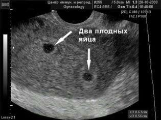
A slight increase in the ovum at a period of 5-6 weeks may indicate that one egg is visualized, but it may well contain two embryos (monochorionic twins, twins). Usually, in this case, a blood test for hCG is done and an ultrasound is repeated a week later to examine both embryos.
Fertile egg too small
The discrepancy in the size of the ovum to the smaller side is an alarming signal. Possible deviations and their causes are presented below.
If the diameter lags behind the growth rate by 2 weeks or more, we can assume:
- fetal malformation;
- frozen pregnancy.
This may be due to early antibiotics, infections, hormonal imbalances, and genetic abnormalities. The forecast in this case is not encouraging. The woman may have a miscarriage or the fetus will have to be removed through an abortion.
If the membranes develop more slowly than the embryo and the growth retardation is more than 14 days, hypoplasia of the ovum is diagnosed. This condition cannot be treated and is an indication for abortion.
If SVD is significantly higher than normal, it suggests the death of the embryo or chromosomal abnormalities of the fetus. This can happen due to the transfer of a pregnant genital infection, in the presence of an abnormal gene or hormonal failure. And in this case, the pregnancy is terminated.
Particular attention is paid to the size of the ovum after the IVF procedure. To control the dynamics of a woman after IVF, an ultrasound is prescribed. Optimal is 28 DPP. At this time, the parameters of the fetus should correspond to 5 obstetric weeks.
Wrong position
Anchoring the embryo below the upper third of the uterus requires careful monitoring by doctors. The low position of the embryo in the early stages does not pose a threat to gestation. There is a great risk that the ovum will rise higher as the uterus grows. However, attachment at the cervix indicates the development of a "cervical" ectopic pregnancy. In this case, the embryo is removed.

Fertile egg of irregular shape
The shape of the ovum is normally oval or round. If it looks flattened from the sides and resembles a bean, this may indicate the tone of the uterus. This condition should be monitored by a doctor. If the patient is not worried about anything, then the deformation has no threat of a pregnancy. With an increased tone of the uterus, doctors take certain measures (taking medications, bed rest) to remove hypertonicity, as well as return the ovum to its normal correct shape. This is achieved by relaxing the muscles of the woman's uterus.

However, if the ovum has an irregular shape, and the patient has pain, discharge or symptoms of cervical dilatation, then urgent measures must be taken. In such cases, a woman is admitted to a hospital for further preservation of the pregnancy.
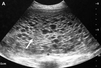
Bubble drift
There is no embryo. There are only overgrown chorionic membranes that form multiple cystic bubbles. They are similar to grape fruits and can be accurately diagnosed using ultrasound. The reason is a poor-quality egg cell, which, when fused with the male reproductive cell, could not provide a full-fledged set of DNA for this process. Papa's chromosomes simply double, and such an embryo, even if it is partially present, cannot grow and develop.
The hCG level is usually significantly higher than the norm, a woman may think that she is carrying twins or triplets.
In every fifth woman, gallbladder drift turns into a malignant formation, prone to rapid and aggressive metastasis - into chorionepithelioma. Therefore, it is important to remove the drift as early as possible - as soon as it is detected.
Empty fetal egg
It should be understood that at very early stages the embryo is not visible in the ovum, and this is the norm. But from about five weeks, the embryo should already be well visualized. In a situation where the embryo is not visible, a second examination is prescribed after one to two weeks. When this time too, the embryo and heartbeat are absent, this indicates anembryony.
You need to know that even in the case of an empty ovum, a pregnancy detection test still shows a positive result. This is due to the fact that certain mechanisms have already been launched in the body, and human chorionic gonadotropin, a special "pregnant hormone", has begun to be produced.
The reasons for the absence of an embryo in the ovum, in most cases, lie at the genetic level. In addition, taking certain medications that are strictly prohibited during pregnancy can cause anembryonia.
When a woman was diagnosed with an empty fetal egg, confirmed by repeated ultrasound, there is no chance of a successful pregnancy this time. The patient undergoes the necessary manipulations.
Symptoms of an empty ovum
An anambryonic anomaly or an empty fertilized egg, the causes of which are still not fully understood, is accompanied by symptoms similar to the course of a normal uterine pregnancy. Already later, when a frozen pregnancy passes into the last stage of spontaneous termination, first minor, and then severe bleeding may occur.
This phenomenon suggests that the body has arbitrarily decided to get rid of pregnancy, and the empty ovum is rejected. In this case, you urgently need to contact a specialist, but you should not panic, because it is much better if the woman's body itself rejects an empty body than if it was necessary to stimulate it with medication or surgically. However, what action should be taken by a woman who suspects she has an empty fertilized egg? HCG, ultrasound and a trip to the doctor to find out the reasons and pass all the necessary tests - this is how you should proceed.
Pregnant women should be especially sensitive to their body and in case of the slightest suspicion immediately consult competent and experienced doctors who will provide an individual, comprehensive treatment. This is the only way to expect a normal pregnancy after an empty ovum in the future.
HCG for anembryonia
With anembryonic disease, the pregnancy test and clinical signs of pregnancy will be positive, since the invasion of the trophoblast into the endometrium promotes the production of chorionic gonadotropin (hCG).
Embryonic treatment
Although anembryonia can result in spontaneous miscarriage, if it is detected, it is necessary to remove the ovum. In the early stages, medical abortion is possible, it is carried out under the mandatory supervision of a doctor. At a later date, curettage of the uterus is done (more details here). The woman is prescribed antibacterial drugs and drugs that reduce the uterus.
It is allowed to become pregnant again after 3-6 months. During this time, the body will recover. It is advisable to plan and prepare for conception. If the next pregnancy also ended unsuccessfully, you need to establish the cause and undergo comprehensive treatment for anembryonia. Both spouses are examined, since the cause of the anomaly may be a genetic defect in the man.
The specialists of the clinic "AltraVita" carry out pregnancy management from an early period. If anembryonic disease is detected, we provide comprehensive treatment. In the future, with us you can be examined and prepare for pregnancy. Even with another frozen pregnancy, there is no need to despair, since in the case of genetic disorders in one of the spouses, IVF can be done with donor material.
Complications
Women who do not go to a gynecologist in a timely manner with signs of pregnancy or their disappearance may face severe complications. If an empty fetal vesicle remains in the uterus for up to 2 weeks or more, an inflammatory reaction occurs, symptoms of acute endometritis appear. The temperature rises, serous-mucous discharge appears from the vagina, signs of intoxication are disturbing - weakness, headache. In the absence of treatment or an untimely beginning, acute inflammation can go to the muscle layer, peri-uterine tissue, and in severe cases lead to peritonitis. If treatment has not been carried out, chronic endometritis develops.
A sign of rejection of the remnants of the embryo from the uterus is the appearance of bloody discharge from the genital tract. But with insufficient uterine contractility, hemostasis disorders, bleeding develops. It cannot end on its own, so medical attention is required. The consequence of bleeding is infection of the uterine cavity, anemia. Complicated by anembryonia, the subsequent habitual miscarriage, secondary infertility.
Pregnancy after anembryonia
Fortunately, after one anembryonic pregnancy, the vast majority of women do not have problems with the onset and gestation of a subsequent pregnancy.
FACT: up to 90% of patients who have had a single pregnancy loss in the past safely carry and give birth to healthy children.
If miscarriages happened 2 or more times, we are already talking about the habitual miscarriage. In this case, the couple needs a more detailed examination and treatment.
Why the fertilized egg is not visible
Despite the fact that an ultrasound scan to view the ovum is recommended as early as the sixth week, it is not uncommon, even at a later date, it cannot always be considered. Moreover, at 12 weeks, in some cases, even the heartbeat is not heard. Doctors say there is no need to panic. Even such a modern and reliable study as ultrasound does not always provide one hundred percent comprehensive information. The lack of a proper and expected result can be affected by many nuances, both technical in nature and related to the human factor. Often, in order to get a complete picture of the course of pregnancy, specialists need to study additional results from studies other than ultrasound. These include laboratory tests and examinations by a gynecologist.
If, during an ultrasound scan, the embryo in the ovum is not visible, the doctor leading the pregnancy will write out a referral for a blood test for hCG (human chorionic gonadotropin). During normal pregnancy, the level of this hormone gradually increases in accordance with the weeks of gestation.
At the same time, if you do not look at the ovum during ultrasound, it is impossible to exclude the possibility that the period was calculated incorrectly. It is possible that it is small enough for the fetus to be viewed on a monitor screen. Doctors calculate the term in weeks, which are called "obstetric". They are counted from the first day of the absence of menstruation. Therefore, calculation errors are common. To establish the exact date, you should re-take the tests and go through the ultrasound again. To do this, you should contact another medical institution.
Doctors pay attention that the exact date when the embryo can be viewed on an ultrasound scan, and not just a separate fetal egg surrounding it, directly depends on the personal characteristics of the organism of the expectant mother, as well as the place of fixation of the egg in the cavity uterus. An important role is also given to modern equipment, with the help of which ultrasound is carried out. For this reason, it is recommended to carry out ultrasound only in competent clinics with modern equipment and experienced specialists.
What to do if the fertilized egg does not meet the standards
Do not panic and worry if the size of the ovum according to the results of your ultrasound differs slightly from the norm. Individual fluctuations downward are quite possible if ovulation was late, implantation took place later than 7-8 days after ovulation, and large sizes may indicate that ovulation happened earlier or early implantation.
Only significant deviations (more than 30% of the norm) can be considered alarming, in this case doctors they will definitely try to find out why the size of the ovum does not correspond to the norm for weeks pregnancy.
Sources of
- https://VseProRebenka.ru/beremennost/plod/malenkoe-plodnoe-yajco.html
- https://MedAboutMe.ru/articles/dolgozhdannaya_beremennost_razmer_plodnogo_yaytsa_po_nede/
- https://o-krohe.ru/beremennost/plod/kak-vyglyadit-plodnoe-yajco/
- https://www.cironline.ru/articles/92422/
- https://www.probirka.org/biblio/polezno/9222-normi-plodnogo-yaytsa-po-nedelyam
- https://o-krohe.ru/beremennost/plod/nedeli/plodnoe-yajco/
- https://www.probirka.org/biblio/polezno/8495-plodnoe-yaytso-i-izmenenie-ego-razmerov
- https://www.probirka.org/zhenskoe-besplodie/9282-pustoe-plodnoe-yaytso-simptomi-i-priznaki
- https://nova-clinic.ru/statyi/anembrioniya/
- https://AltraVita-IVF.ru/informatsiya-dlya-patsientov/spravochnik-zabolevanij/anembrioniya.html
- https://www.KrasotaiMedicina.ru/diseases/zabolevanija_gynaecology/anembryonic-pregnancy
- https://www.altermed.ru/gynecology/article/anembrioniya-animaonline-pregnancy/
- https://www.medar.kz/kogda-mozhno-uvidet-plodnoe-yajco-na-uzi.html