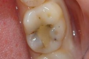
 Treatment of carious foci by the filling method does not always guarantee the final disposal of the problem. Often, caries manifests itself after some time( usually in 2-4 years) after dental intervention.
As the inflammatory process occurs under the seal, it is difficult to identify it in the initial stages and often a person turns to the doctor when the secondary caries manifests itself with bright symptoms.
Content
- Secondary VS recurrent - sort out the terminology
- Development Mechanism
- bacteria returned to renew their "rotten" deal
- diagnostic methods
- Clinical picture
- dental care
- Repeated filling
- adhesive restoration
- microprosthetics
- crowns Installation
- Possible consequences
- Preventive measures
Secondary VS recurrent - we will understand in terminology
Under the secondary caries we mean the imageThe development of new carious foci under a previously sealed tooth, which was provoked by bacteria that penetrated through microcracks between the hard tooth tissue and the filling. Also, this term means spreading the lesion to a healthy enamel near the seal.
In the case of recurrent caries, it is a resumption of the carious process at the site of treatment. As a rule, the cause of the pathology is the negligence of the dentist:
- the affected tooth cavity was not completely processed( there are remains of decomposed dentin);
- antiseptics have not been used( the infection makes itself felt even in the presence of the slightest bacterium).
However, it is not possible to identify with certainty the cause of the reoccurrence of the carious process: whether this is caused by a doctor's mistake when treating the affected cavity, or whether the caries is a consequence of the shrinkage of the filling material. In most cases, these processes are manifested equally often.
In addition, both secondary and recurrent caries can develop simultaneously.
Developmental mechanism
Secondary carious lesion affects healthy solid tooth tissues that are close to the established seal. The 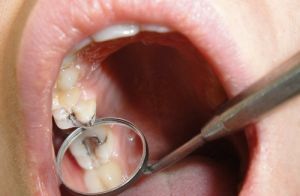 disease progresses in stages:
- in the first stage of the , microcracks form between the filling material and the dental tissue;
- in the second stage of , bacteria and other pathogens penetrate into the formed cracks;
- in the third stage of , active reproduction of bacteria is observed, which in the course of their vital activity produce acids that adversely affect the state of the enamel and fillings, as a result, the composite begins to tear away from the tooth.
Bacteria return to resume their "rotten" case
The main cause of caries formation under an earlier established seal is the initially incorrect treatment of the damaged tooth:
- All lesions of were not removed when cleaning the carious cavity.
- Preparation of the outer surface of the tooth before sealing was not performed properly .As a result, the tooth tissues adjacent to the filling begin to crumble over time. On the place of the formed cracks new foci of defeat will arise.
- Features of shrinkage of a seal .In some cases, the filling material can sag, provoking an aggravation of the secondary destruction of the already affected cavity. A similar defect occurs in light-polymer models, the size of which can vary under the influence of light. As a result, cracks appear between the material and the dental tissues, through which various pathogens penetrate.
The following factors affect the wear resistance:
- simultaneous effect on tooth enamel low and high temperatures - a similar phenomenon is possible with the use of hot dishes and drinks with ice;
- abuse of solid food;
- pathology of the jaw structure( their incorrect closure, bite problems);
- bruxism - grinding, tapping and clenching of teeth promotes active friction and thinning of tooth enamel;
- neglect of hygiene rules, untimely brushing of teeth, lack of dental floss in the oral cavity allows food residues to gather between the teeth, which creates favorable conditions for the formation of bacterial plaque.
Diagnostic methods
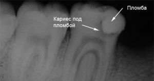
How cavities under the seal look on the X-ray photo-image
A standard examination helps to identify the existing pathology of the dental tissue. However, in some cases, the diagnosis requires the use of the viziograph.
Visigraphy is a relatively new diagnostic method that allows a full assessment of the condition of the teeth and gums and the selection of the appropriate treatment package. Indications for such a diagnosis are complicated caries, periodontitis and pulpitis.
This method of examination is also used when examining teeth with previously installed seals. Among the advantages of this type of diagnostics are the following:
- Operationality .The picture is taken in a short time.
- Security .In comparison with the classical X-ray, the radiation dose is 10-15 times less.
- Sharpness and informative image .The image is sent to the computer monitor, which allows you to view all the available foci of inflammation in the mouth.
The use of visionography is appropriate not only in diagnosis, but also in the treatment of secondary carious lesions, which makes it possible to evaluate the effectiveness of restoring a damaged tooth.
Features of the clinical picture
For secondary caries, the following symptoms are typical:
- pain in the teeth and jaw, intensifying when touched;

- gum bleeding;
- inflammation and swelling under the seal;
- is an unpleasant, putrid odor from the mouth;
- redness of the oral mucosa;
- formation of black dots on enamel of healthy teeth;
- seal instability;
- sensitivity of tooth enamel;
- color change of the sealed material.
The insidiousness of caries, which develops under the seal, is its initially asymptomatic course. Acute pain occurs even with neglected pathology, the treatment of which often requires radical dental procedures.
Rendering of dental care
Treatment depends on the degree and severity of the secondary caries. After a complete diagnosis, the dentist chooses a specific method of therapy, or completely removes the tooth, if his deep layers were damaged along with the root.
Re-sealing
The procedure of re-sealing is carried out in several stages: 
- reamers the damaged part of the tooth;
- removes damaged or dead teeth and the rest of the seal;
- cleans enamel and dentin;
- treat the tooth cavity with antiseptics;
- is laying an insulating gasket;
- is installing a new seal.
To ensure the effectiveness of the procedure, the carious areas are stained with a special solution. By the results of the reaction, it becomes clear whether all the damaged tissues have been removed.
Adhesive restoration
This method of treatment is an alternative to the installation of a dental crown. The main advantage of adhesive restoration is the minimal impact on the tooth enamel, while achieving high integrity of the tooth itself.
The procedure is as simple as possible. The prepared tooth is treated with an adhesive polymer, the main task of which is to restore the destroyed enamel and reduce its sensitivity to different temperature effects. The treatment is reliable and durable, and as a result, the patient gets an attractive smile.
Microprosthetics
To date, microprosthetics is considered to be a progressive and effective way of treating a caries-affected tooth, even in small patients.
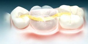 The technology implies the installation of special ceramic( or other materials) tabs-prostheses. A cast of tooth, affected by caries, is made, its edges are processed and sent to the laboratory to create suitable tabs.
The design fully corresponds to the shape, size and color of the tooth, which makes its presence invisible to others. In addition, due to the tight fixation, such a tab is not felt by a foreign body in the mouth. If it is not possible to perform the procedure, a re-installation of the crown is used to restore the tooth.
Installing the crown
The crown is installed provided it is not possible to fill, build, and other methods of treatment. However, crown installation is possible in the absence of damage to the dental root.
For the production of crowns, alloys are used from medical steel( because of their unaesthetic appearance, a similar method is used in the treatment of distant teeth), cermets( characterized by strength and attractive appearance) and ceramics( most similar to natural teeth, but unable to cope with strongloads).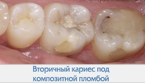
Preparation for the procedure includes the removal of the deadened tooth or its turning and subsequent grinding. After this, a plaster cast is made, the basis of which will serve to create a future crown. If the crown is manufactured for a long time, the patient is provided with an auxiliary plastic structure that will protect the turned tooth.
Initially, the crown is installed on a temporary cement slurry so that the behavior of the affected tooth can be traced to adjacent healthy teeth. In the event that no adverse effect was observed, the product is fixed to a permanent cement.
Possible consequences of
Untimely treatment is fraught with the development of a number of complications:
- damage to bone tissue down to its deep layers;
- destruction of the canal and root of the tooth;
- development of the disease on adjacent healthy teeth;
- complete tooth decay.
In secondary caries, the inflammatory processes in the pulp and its subsequent necrosis are especially dangerous, which can be caused by traumatic preparation, the use of irritants in the treatment of the tooth cavity, the toxic effect of the filling material.
Preventive measures
 As a preventive measure, it is recommended not to neglect daily oral hygiene. To do this, use certified toothpastes, antiseptic rinse, dental floss.
It is also important to remember the timely visits to the doctor. Ideally, a visit to the dental office should be done twice a year, especially if there are previously established seals or dental crowns.
This will detect the appearance of caries in the early stages and prevent the progress of the disease.
