 Teeth are important bone formations located in the oral cavity of a person. They perform the function of primary mechanical processing of food, and also help in the pronunciation of sounds and the formation of speech.
Teeth are arranged in an orderly manner on the lower and upper jaws in the dentition, they have a definite shape and structure. Are able to get food due to the inside of blood vessels, and thanks to the nerves - to perceive pain and other irritations.
Throughout a person's life, only temporary replacement of temporary fragile teeth( dairy) with firm constants takes place.
Contents of
- Each tooth has its name and place in the mouth
- Anatomical structure of
- Histological structure of
- Differences of dairy and permanent teeth
Each tooth has its name and place in the mouth
Depending on location and functional role, all teeth are divided into four subgroups:
- central and lateral incisors;
- fangs;
- small chewing or premolars;
- large chewing or molars( including wisdom teeth).
The upper and lower jaws have a symmetrical structure and include rows with the same number of teeth from each subgroup.
The functional load is the following:
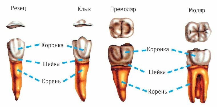
- The teeth in the front part are called by the cutters. There are only eight of them - four on the bottom and top. The largest are the two upper incisors. All teeth of this subgroup have a flattened appearance with sharp edges and resemble a chisel in shape. Their main purpose is biting food and dividing it into fragments.
- There is one canine on both sides of the incisors. This name was obtained because of the existing anatomical similarity with the teeth of predatory animals. Fangs during the process of eating help to divide it into small pieces. The main task of chewing teeth is thorough grinding of food.
- premolars have a convex crown in the form of a prism. On the upper part there are two tubercles, between which there is a furrow. Molars are considered the largest teeth. Their chewing surface has the form of a quadrilateral with convex tubercles separated by fissure. There are eight molars and premolars in total.
At the age of 16 to 26 years, young people can cut the eighth tooth in a row. He is also referred to as molars, and popularly called wisdom teeth. There are times when, due to the lack of sufficient space on the jawbone, they cut through incorrectly and take an unnatural position in their row.
Anatomical structure of
From the point of view of anatomy, a person's tooth is conditionally divided into three parts:
- The crown of the is the visible part of the tooth, located above the gum.
- With the of the root, the tooth is retained qualitatively in the alveolus - a peculiar jaw groove. Roots can have one or more shoots, depending on the type of teeth.
- Between the crown and the root is the neck of the tooth. It is distinguished by a narrowed shape and is closed on all sides by the edges of the gum.
The inside of the tooth has a cavity. In the part of the crown, it completely repeats its shape, but at the root it continues in the form of its channels and ends at the apex with an opening. The wall of the tooth cavity is usually called a vault, and the cavity at the beginning of the root canals is called the bottom.
Strong connective fibers hold the tooth root and neck with bone jaw tissue. Near the neck they are located almost horizontally. This gives them the opportunity, together with the gum and periosteum, to fix the tooth from all sides and hide the root from the external environment.
In addition to fixation, the ligament is assigned the role of a kind of shock absorber. After all, in the process of chewing food on the teeth is a significant load, which, without connective fibers, could lead to injuries to the bottom of the dental hole.
Histological structure of
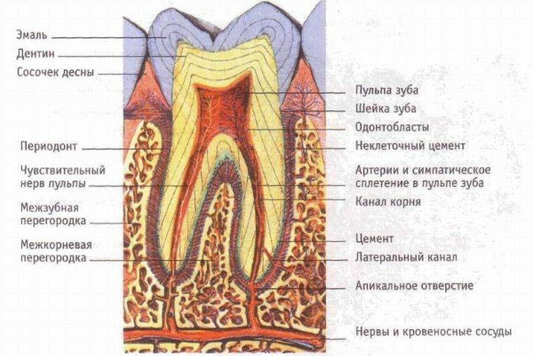
The tooth contains several types of tissues, but dentin is its main component. Above the dentin in the root part of the tooth is covered with a thin layer of cement, and the crowns with enamel.
Enamel is a surface coating of the tooth, which has a high wear resistance. It serves as a "cover" protecting the open part of the tooth from external factors.
This coating is considered the most solid substance in the human body. This is due to the content in it of a large number of minerals.
Reasons for thinning of enamel can be:
- insufficient amount of minerals in the body( calcium or phosphorus);
- regular use of sweets and carbonated drinks;
- mechanical action( cracking of nuts, opening of bottles with bottles, etc.)
- cleaning of teeth with rigid brushes.
Damage is often caused by bacteria in the oral cavity, which leads to the development of a disease such as caries. 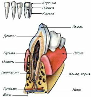
Holistic tooth enamel does not allow a person to feel discomfort or pain during eating.
Dentin is the basis of the tooth. In its composition, it has a similarity to bone tissues, but has increased strength due to the high content of inorganic substances. The tissue of dentin is penetrated by a large number of very thin channels. Due to this, metabolic processes are actively occurring in it, ensuring the vital activity and development of the tooth.
The nutrition of the tooth is placed on a soft pulp, located in its entire cavity. In its form it is completely identical to the appearance of the tooth and is a tissue with an abundance of blood and lymphatic vessels, nerves and cells of odontoblasts, the processes of which are connected with the thin canals of dentin.
Depending on its location, the pulp can be root or coronal.
The main functions of this soft fabric are:
- dentin formation;
- providing dentin with nutrients;
- transmission of information about pain stimuli through sensory nerves;
- removal of dead cells and destruction of foreign microorganisms.
The neck and root of the tooth cover a peculiar tissue, which in its composition resembles bone, and is called cement. Thanks to it, the tooth is connected to the ligamentous apparatus. In addition, cement is required for:
- protection of the dental root from external factors;
- protects dentin from penetration of microorganisms provoking caries;
- tight fixation of the tooth in the alveolus.
The presence of dental lymph vessels makes it possible to carry out the outflow of lymph through the nearby lymph nodes. Some of them are amenable to palpation, and, consequently, their examination may indicate the presence of inflammatory processes.
How are the human teeth:
Differences between dairy and permanent teeth
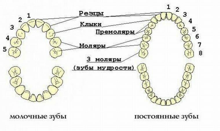
At the age of 6 - 9 months, the first dairy teeth begin to appear in babies. Usually the whole dentition is formed by three years. The total number of temporary teeth is 20.
By the age of 5 to 6, the dairy begins to fall out, and permanent teeth appear in their place. Temporal and molar teeth are similar in structure, but they have a number of significant differences:
- The size of the crown is much smaller in the first.
- Enamel is thinner than , and dentin contains a small amount of minerals. For this reason, children often develop caries. Incisors more convex .And the roots are small and less strong. Therefore, in childhood, tooth replacement is considered a virtually painless process.
Despite sufficient strength, a person's teeth are considered to be a system in the body that can easily undergo the loss of full or partial functionality under the influence of various factors.
In addition, the teeth are not capable of regeneration, so they require care and careful attitude throughout life, from an early age.
