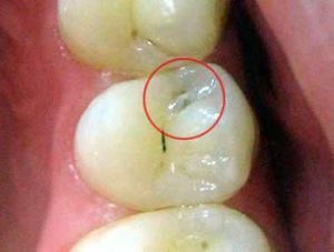
 Hidden caries are called imperceptible tooth decay. In the medical literature this term does not occur, but recently practicing dentists use it more and more often.
You can detect the disease only with the help of special diagnostic methods. Severely detectable caries can develop in those parts of the tooth where food remains, for example, in the interdental space.
The insidious feature of the latent form of caries is that a person can not guess at the development of this disease.
Contents of
- Where can pathogenic bacteria escape?
- Factors provocateurs
- Symptoms and clinic
- Diagnostic criteria
- Methods of treatment
- Possible consequences
- Preventive measures
Where can disease pathogens hide?
Depending on the place of occurrence, inconspicuous caries are divided into the following types:
- Contact .It spreads between the teeth. The person notices the presence of the disease when she has already hit the entire tooth and there was a sharp pain. But the dentist can easily identify the disease at an early stage. Most often, the destruction begins because of the constant presence in the interdental space of food particles. The main disadvantage of contact caries is that its treatment is a rather unpleasant process.
- Fissured .It affects fissures( natural dental cavities).It looks like a small dark speck on the enamel. If such a spot is noticed, it is necessary to urgently appeal to the dentist, otherwise the disease will quickly progress and the darkened enamel will break away, there will be a strong toothache. Fissure caries is usually easy to treat.
- Caries of the tooth root .This type of disease is the most dangerous and problematic. Most often it is diagnosed already with extensive damage not only to the tooth root, but the entire tooth. This disease usually develops in parallel with periodontitis. The root of the tooth is separated from the gum, exposed and begins to deteriorate. To cure this form of the disease is very difficult, in most cases the removal of a sick tooth is inevitable.
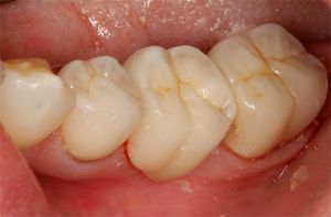
- Secondary .In this case, caries occurs under the seal. It is detected after the appearance of painful sensations or loss of a seal( with a strong growth of the lesion).The initial stage of the spread of the disease can be identified during a preventive examination.

In a photo of a variety of hidden areas in caries: 1-pin, 2-secondary, 3-inside the tooth
Factors provocateurs
The reasons that provoke latent caries can be different. The most basic - non-compliance with oral hygiene, consumption of a large amount of sweet food, gastrointestinal disease, lack of vitamins, decreased immunity, improper formation of teeth, lack of calcium, phosphorus and fluorine in drinking water.
All these factors contribute to poor-quality cleaning of teeth from stones, reproduction of pathogenic bacteria in the mouth, hypoplasia of the enamel. Because of this, hidden cavities arise.
Symptomatology and clinic
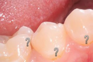 The danger of caries-partisans is that at the initial stages of spreading it does not manifest itself in any way. A person does not feel pain, can not visually( using a mirror) determine the presence of the disease. The latent form of the carious process is manifested after significant damage to the tooth or its root.
Seizure is the defining symptom. This means that the carious opening has increased significantly. Also on the surface of the enamel can be observed dark and light spots.
With extensive damage, a person begins to feel severe pain due to temperature( cold or hot drink, cold air) and chemical irritants( sweet or salty foods).
If the pain arises arbitrarily, this indicates that the caries has developed into pulpitis( inflammation of the internal tissue of the tooth).
Pain can also occur with mechanical pressure on the affected area. Usually this happens during chewing food. The food penetrates into the formed hole and provokes clamping of the vascular-neural bundle( pulp).These sensations can be eliminated by removing food particles from the affected cavity.
Diagnostic criteria
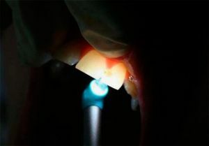
Laser - modern diagnostic method
A latent carious cavity in the tooth is easily detected using modern diagnostic methods. Localization and development stage are practically irrelevant.
However, before using these methods, the doctor must identify the carious process. This does not always happen, since the visual tooth looks healthy, the patient does not complain about anything. For this reason, the carious process remains unnoticed and continues to develop rapidly.
Most often, the latent form of the disease progresses together with other carious processes, the signs of which are visible to the naked eye.
If the patient experiences multiple injuries, then an experienced specialist will perform a thorough examination for imperceptible lesions.
The following diagnostic methods are available:
- Laser .This method is one of the most reliable. It is carried out with the help of a special device - the Diagnostent. If there is a lesion, the device detects it by reflecting the laser beam from the damaged organ and emits a characteristic sound.
- Radiography .X-ray helps identify caries in hard-to-reach areas of the tooth. But the disease must be already sufficiently developed, otherwise the radiography will not reveal it.
- Inspection using the mirror. Visual inspection is the first stage of diagnosis. In this way, the dentist determines whether there is a chance of hitting a certain tooth, and then applies other methods.
- Transillumination of .A bright light is sent to the tooth roots, which illuminates the damaged areas of the tooth. With this method of diagnosis, dentists use a photopolymerization lamp.
Treatment methods
 Treatment of latent caries in general terms does not differ from other forms of the disease and consists in the removal of diseased tissues and tooth filling. If the enamel is damaged or a spot is visible on the tooth, the tooth tissue must be opened. To restore the enamel, fluoride and calcium preparations are prescribed.
If tooth decay develops under the seal, the affected areas are cleaned and the seal is reinstalled. Before the filling, the doctor performs local anesthesia.
With the defeat of the tooth, excision of patients with dental tissues is carried out, then the crown is installed. Another method of treatment is complete tooth extraction and implant placement.
Case from a doctor's practice:
Typical situation: between two damaged teeth is one that does not have obvious signs of caries. But it is likely that a healthy looking tooth can be infected.
So it turned out: the radiograph showed extensive damage to the back wall of the dental tissue. The pulp was not affected, but the bone substance required purification and subsequent sealing.
Karim Pavlovich
Possible consequences of
Caries can lead to various complications:
- allergic reactions to infection;
- occurrence of inflammatory processes in the gums and facial tissues;
- negative impact of the infected tooth on the whole organism;
- occurrence of flux;
- development of the cyst of the jaw.
Because of pain, the food is chewed badly, which is very harmful for the gastrointestinal tract. 
If you do not treat the disease, the consequences can be irreversible and you will have to remove the affected organ. Loss of even one tooth is a great stress for the whole organism.
In addition, such dangerous diseases as pulpitis and periodontitis can develop. Pulpitis is expressed by acute painful sensations when nibbling. Pathology can occur even if caries has been treated. This may be due to the fact that the infected tissue was not completely removed.
Periodontitis is a serious complication that affects not only neural bundles, but also ligaments. If you do not immediately start treatment, the ailment can go into a chronic form.
Preventive measures
To prevent disease, the following recommendations should be followed:
- Regularly brush the teeth of .Do brushing your teeth at least twice a day. During the process,
 should pay special attention to the interdental spaces and other hard-to-reach surfaces. To clean it was more effective, pick up a suitable toothbrush and toothpaste.
 - Drink water that contains a sufficient amount of fluoride .A lack of fluoride can lead to the gradual destruction of enamel. To avoid this, drinking water must be fluorinated. But there is an easier way - the introduction of seafood into the diet. You can also use special rinse aid.
- Do not take too hot or cold food .This can provoke the appearance on the enamel of microscopic cracks. Carious microorganisms can freely enter into them.
- Visit the dentist at least twice a year .Preventive examinations are extremely necessary. Adults need to spend them every six months, children - 4 times a year. This will help prevent various diseases of the oral cavity, as well as quickly and without consequences cure already available. If plaque accumulates on the teeth, professional cleaning should be carried out to stop the spread of bacteria.
