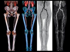
 Angiography is an x-ray method for diagnosing various diseases of veins and arteries, which allows to assess their general condition, as well as the condition of the walls and the presence of disturbances in the circulation.
The essence of the diagnosis is that a special contrast agent is injected into the vessels, after which several photographs are taken. As a result, it gives the right to confirm or deny a particular disease, including the appointment of the right method of treating the patient.
Contents
- Indications for diagnosis
- When the procedure is not applied
- Preparing for the
- procedure How the
- is being performed Decoding of the results
- From the practice of the
- patients Price of the question
Indications for the diagnosis
The main indications for the appointment of this procedure are diseases or pathological changes of the arteries, in particular:
- atherosclerosis;
- thromboembolism;
- diverticulitis;
- aneurysm and others.
In addition, angiography of the vessels of the lower extremities is prescribed to the patient for various diseases of the veins, namely: 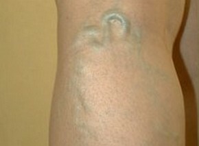
- phlebitis;
- varicose veins;
- trophic ulcer;
- thrombophlebitis.
As for other diseases, the procedure is often prescribed for such diseases:
- diabetic foot syndrome;
- diabetes mellitus;
- purulent-necrotic lesion of the extremities.
In practice, most often there is a need for angiography of the lower extremities. This is more associated with a large number of diseases of the arteries and veins.
Angiography of the upper limbs is mostly performed in the event of various injuries. With venous disease, it is customary to prescribe phlebography. If we are talking about diseases of the arteries - arteriography.
In the process of diagnosing arteries of the lower limbs, the catheterization method is used. In order to assess the condition of the arteries of the foot, a tibial tibial puncture is used.
When the technique is not used
 Vessel diagnosis is a procedure in which iodine-containing medications are used. Quite often it is possible to meet patients who have personal intolerance to such drugs. Therefore often such medicines are changed to non-invasive.
Patients who have certain diseases can not be admitted to this procedure. In particular, we are talking about such diseases:
- chronic heart disease;
- kidney failure.
It should be noted that for the female, the main contraindications are considered to be pregnancy, since X-rays can cause irreparable harm to the health of the baby's future.
Preparation for
procedure This research technique is rightfully considered an invasive procedure, which, first of all, requires a certain preparation.
As a rule, doctors categorically prohibit the use of alcohol not earlier than 10 days before the diagnosis. Moreover, 5 days before the study, it is necessary to completely exclude the use of medications, whose actions are aimed at diluting the blood in the body.
On an individual basis, treating physicians may additionally prior to the procedure prescribe ultrasound and fluorography.
In addition to the basic training procedures, additional ones are also assigned, which include:
- no later than a day is done a test for the allergic reaction of contrast medium, for this patient, no more than 2 ml of the drug is administered and the reaction is followed;
- in the place where the catheter will be inserted, shave the hair;
- a few hours before the procedure is performed cleansing of the intestine, this is due to the fact that after the study for some time you can not get out of bed;
- for a category of patients who have kidney problems, drink plenty of water;
- a few minutes before the start of the study, measure the pressure;
- , most patients are asked to take a sedative so that the pressure is normal;
- for 10 hours before the study can not eat;
- during the procedure on the body of the patient should not be metal objects, so they must be removed in advance.
How the
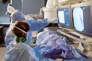
is being conducted The average study is no more than 20 minutes. The place where the injection will be made is treated with a special antiseptic drug.
After this, the specialist uses a special needle to puncture the necessary vessel. In the future, the needle is removed and a contrast agent is introduced through the catheter.
At the end of the injection, aiming or layered shots are taken. At the end of the procedure, a catheter is withdrawn from the patient, and a special dressing is used to stop bleeding.
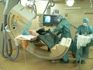 It is worth noting that at the end of the procedure, the patient should still be under the supervision of specialists for some time in order to avoid any consequences.
The procedure is performed under local anesthesia, so all painful sensations are completely excluded.
When entering a contrast agent, there may be a feeling of heat in the oral cavity, which is a normal reaction to the body and panic in this case is not worth it.
Decoding results of
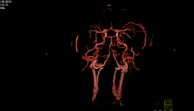
Based on the results of the images, the specialist begins to decipher the obtained data, in particular, the following parameters are investigated:
- blood flow in the vessels;
- presence of clot formations, including aneurysms;
- degree of closure of the lumen of blood vessels due to blood clots;
- propensity to angiospasm;
- in the study of neoplasms, determines the presence of blood circulation in them;
- the presence of possible hemorrhages, including the integrity of the bloodstream.
The entire decryption procedure takes no more than 15 minutes, after which the patient is transferred the results. It is also possible to record results on an electronic medium.
If the patient is on inpatient treatment, the results are transmitted directly to the treating physician.
If the patient has performed the diagnosis on his own initiative, he should in the near future consult his doctor for advice.
If any abnormalities in the operation of the vessels are revealed, a necessary course of treatment will be appointed, which will allow to get rid of this or that disease in a short time.
From the practice of patients
Reviews of patients who underwent angiography of vessels of the lower extremities.
Hello! My name is Marina. I would like to tell you how I underwent angiography. At me varicose phlebectasia and to me have appointed or nominated similar diagnostics.
I want to say at once that I spent 3 days in the hospital - a day before the procedure and two after. During the diagnosis, I did not feel pain, but some discomfort was present. The study took no more than 30 minutes of time. The only drawback is that after that you can not get up during the day.
When I was told about the need for this procedure, I was very frightened, but it turned out all wrong and scary. At the puncture site, I had a bandage and after 2 days I was removed. In general, there is nothing wrong with that.
Marina
Good afternoon! I hasten to share my joy. My name is Alina, I had health problems( varicose veins), but now everything has passed. The diagnosis was helped by angiography.
At one time, when the doctor told me about the need for this study, I refused, but as it turned out in vain. Diagnosis took me 25 minutes, I did not feel pain.
The only drawback for me was the burning sensation in the mouth when entering contrast medium, as well as the ban on lifting from the bed during the next day. It was uncomfortable with the bandage, which was imposed to stop bleeding( a little crushed, because of what was a small puffiness).
In the hospital, they treated me very well, carefully and carefully. Thanks to specialists, thanks to whom I was prescribed a course of treatment, thanks to which I got rid of my disease.
Alina
Price
The price of such a diagnosis depends largely on the level of the medical center, as well as the equipment with which it is performed.
The average price of angiography of vessels varies from 11 000 to 15 000 rubles. This cost includes all the necessary drugs, including a contrast agent.
