 Among all diseases of venous vessels, the most dangerous are pathological disorders that develop in the basin of the inferior vena cava.
The collapse of this largest vessel, which collects blood from the lower parts of the body, leads to severe consequences associated with the onset of chronic insufficiency of the venous apparatus and a violation of the function of internal organs.
The syndrome of compression of the inferior vena cava is often identified with the thrombosis of this vessel, because it is a consequence or complication of it. Why is this situation developing, how to deal with it, and whether such a reversal of events can be avoided?
Contents
- Anatomical and physiological reference
- Where does the problem come from
- How does the violation occur
- Diagnosis and treatment guidelines
- Consequences up to death
- The danger that is better to prevent
Anatomical and physiological information
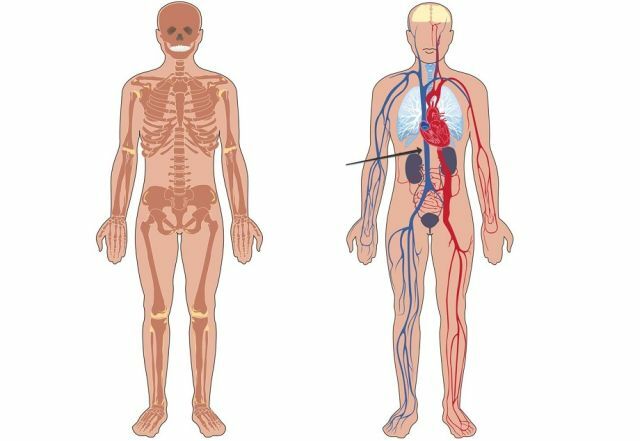
The lower hollow vein( NIP) is one of the largest andsignificant blood vessels of the circulatory system. It has its own pool of a ramified network and collects blood from the entire lower body floor.
This vein is located on the right side of the aorta in the retroperitoneal space( the area of the fiber from the diaphragm to the small pelvis).Within the abdominal cavity, the LIP passes behind the small intestine and pancreas, and runs into the transverse groove of the liver.
Further, the vein penetrates through the diaphragm into the middle part of the mediastinum, where it transfers blood directly into the cavity of the right atrium. There are no valves in the NIP, its diameter varies depending on the respiratory cycle from 21 to 35 mm( it is wider on exhalation than on inspiration).

The photo shows the lower floor of the vein
The lower vena cava basin is the most developed and strong system of venous vessels in the human body( contains approximately 65% - 70% of all venous blood).This network is formed by vessels of different calibers. The NIP has several tributaries. Some of them are internal:
- renal venous vessels;
- veins of the ovaries and testes;
- vein of the liver;
- adrenal branches.
Another part of the tributaries is the parietal vessels:
- veins of the diaphragm;
- vessels of the lumbar region;
- veins of the buttocks( upper and lower);
- lateral-sacral;
- ileal tributaries.
What is the inferior vena cava, its functions and anatomy are detailed in the video:
Where does the problem come from
? The syndrome of the inferior vena cava is a symptom complex that develops as a result of complete blockage( occlusion) or partial obstruction( parietal thrombus) of the main venous trunk collecting bloodfrom the lower extremities, the organs of the abdominal cavity and pelvis.
There are 2 forms of this pathological condition: acute thrombosis of the inferior vena cava and chronic obstructive obstruction.
The main cause of occlusion of a large vessel is ileofemoral thrombosis( at the level of the iliac and femoral veins), which tend to increase ascending. Most often the syndrome of the inferior vena cava happens in the following situations:
- on the background of pregnancy , especially the prolific or large fetus;

- in the presence of tumors of the abdominal cavity( liver), retroperitoneal space( kidneys, pancreas) or pelvic floor( urogenital system);
- for retroperitoneal fibrosis ( Ormond's disease) - mechanical compression of vessels with sclerotically altered fatty tissue;
- congenital atresia of the lumen of the ;
- thrombosis and occlusion of the hepatic veins of congenital or acquired origin( Bada-Chiari syndrome).
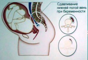
The first cause( pregnancy) is the most common. In later terms, the growing uterus always compresses the lower vena cava to some extent, which is manifested by increased venous pressure in the legs and a decrease in the return of blood to the heart.
Reduction of cardiac output and blood volume in a small circle of blood circulation leads to oxygen starvation of the whole organism. However, the clinic of the syndrome of compression of the inferior vena cava develops only in 10% of pregnant women, in others the venous outflow is performed through a network of collaterals( bypasses) that are formed.
How the violation of
is manifested The severity of the clinical picture of the syndrome of the inferior vena cava depends on the level of occlusion or compression of the venous trunk. When overlapping the lumen above the site of the exit of renal vessels, renal damage develops( nephrotic syndrome with edema and increased protein content in the urine) with further increase in kidney failure.
If the lesion level is below the renal veins, then the lower extremities( varicose veins, numerous trophic ulcers of the lower leg, numbness and swelling of the legs) suffer.
 Pain in this pathology is more common - legs, inguinal area, lower back with buttocks and abdomen.
Puffiness is expressed - it usually covers the legs all the way from the groin to the toes, as well as the genitals and the anterior part of the abdominal wall. The enlarged subcutaneous veins are most visible on the shins, less on the hips, strongly visible on the abdomen - along the sides of the abdominal wall and above the womb, at the top they connect to the superficial veins of the thorax.
Features of the syndrome of the inferior vena cava in pregnant women are due to the fact that the expressed compression of this vessel with a large uterus( after 25-26 weeks) reduces uterine and renal blood flow, which adversely affects the development and condition of the fetus.
This is especially evident in the position of the woman on the back - there is a sudden weakness, dizziness, choking, low blood pressure until fainting.
The future mother reduces glomerular filtration and other kidney functions, sudden premature plaquent detachment and even rupture of the uterus may occur. Such women often have signs of varicose disease and hemorrhoids.
Diagnosis and treatment guidelines for
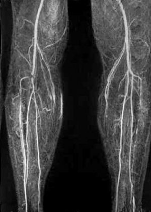
If you suspect a development of NIP syndrome, emergency medical attention is necessary. Diagnostic measures consist in carrying out phlebography with a contrast substance, on pictures it is possible to establish localization of narrowing or blockage of a vein. To complete the  diagnosis, additional ultrasound of the vessels, as well as MRI, is performed. Laboratory diagnostics consists in carrying out general and biochemical analyzes of the urinary residue and blood, research of the coagulation system. The choice of an individual treatment regimen is appointed after evaluation of the results of the study.
Treatment can be radical operational and conservative, the latter given preference. The goal of drug therapy is to eliminate the pathological process and restore normal blood flow.
Individual doses of thrombolytics and anticoagulants( for dilution of blood and removal of thrombi), if necessary, non-steroidal anti-inflammatory drugs, sometimes require antibacterial medications according to indications are selected for this purpose.
In addition, compression therapy, balneological and physiotherapy procedures are used.
Surgical intervention physicians practice in rare cases, for example, with mass formation of thrombi in the lower limbs or when narrowing the lumen of the LEL above the location of the renal arteries. Effective is the method of autovenous bypass, less often - prosthesis of the inferior vena cava.
Consequences up to the lethal outcome of
The most formidable complication is thrombosis in the inferior vena cava system, it is about 10% of the total number of thromboses.
Most often, this condition develops ascending hematogenous way from veins of smaller diameter or as a result of compression of the vessel with a tumor.
The clinical picture of acute thrombosis of LIP depends on the speed of blood clots, the degree of occlusion of the lumen of the main venous trunk and its tributaries, and also on the compensatory force of the collateral pathways. The worst prognosis is associated with the rapid progression of thrombus and the development of pulmonary embolism.
 The rapid development of the occlusal obstruction of the inferior vena cava is manifested by severe pains in the lower abdomen and back, swelling and cyanosis of the limb, spread over the entire area of the extent of thrombosis.
Blocking of the NIP in the area of the exit of the renal veins contributes to the onset of severe consequences, for example, damage to the parenchyma of the organ with the development of renal failure.
Occlusion in the zone of the hepatic vessels leads to a violation of the basic functions of the liver and to thrombosis of the lumen of the portal vein, which significantly burdens the prognosis.
With this localization, abdominal pain, especially on the right under the rib and in the epigastrium, increases in the liver and spleen, abdominal hydrops, a characteristic appearance of the enlarged veins pattern on the anterior surface of the abdominal wall, similar to the head of the jellyfish.
Danger, which is better to prevent
Preventive measures are based on the etiology of the pathological condition.
The task is to prevent the occurrence of a reason for the development of such a violation. Recommendations:
- monitoring of the blood coagulation system, especially if there is a hereditary predisposition to increased thrombosis;

- Taking Aspirin Drugs at a dose of 50-75 mg every day after consulting a doctor;
- at the first symptoms of trouble in the body to seek help from the appropriate specialist;
- if you are pregnant for more than 26 weeks, you can not rest or exercise in the "lying on your back" position;
- recommended regular exercise( walking, swimming);
- in the process of labor is preferred position with a high head or sitting.
With a timely diagnosis of the problem and proper adherence to the recommendations of doctors, the prognosis for NIP syndrome can be quite encouraging.
