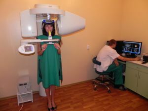
 For a long time, medicine has become stagnant, because, in fact, it was very difficult to determine whether a person is sick or not. Sometimes even operations were performed on quite healthy people, due to false symptoms, the so-called phantom pains.
This was all due to the fact that a person could be scared, and the symptoms that seemed to him, he had, were nothing more than a placebo effect.
And the doctor himself could not assess the patients' internal state in any way. And this continued until the Roentgen invented the very rays from which the revolution began.
At first, only the bones showed through to the person, but eventually the scientists learned to use ultrasonic waves, which changed the face of medicine forever. But so far, the invention of X-ray is incredibly in demand, and one of the devices in which it is used to this day is the tomograph.
Contents
- The concept of the tomography of the teeth
- What exactly can reveal a three-dimensional snapshot of the teeth?
- How it works
- Variety of research
- Contraindications and indications for the use of the
- method Word for the patients
- Advantages and disadvantages
- Price of the question
The concept of the tomography of the teeth
Computed tomography of teeth is an incredibly important milestone in the development of modern medicine. This revolutionary method allows you to directly view the state of the teeth, using X-rays.
In addition to a 3D snapshot of a single tooth, there is a full procedure for the entire oral cavity, called a tomography of the jaw.
CT images taken in three-dimensional format, allow doctors to fully assess the entire state of the jaw, as well as clearly identify where the hearth of a disease, so that in the future it was easier to eliminate it.
That's why, absolutely all operations performed on the teeth can be started only after a full examination and tomography of the jaw. 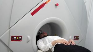
With the help of a tomography, a patient can learn not only about the general condition of his teeth, but even about the state of the jaw bone tissue, and even assess the condition of the maxillary sinuses, if necessary.
Thanks to all these features, this procedure was an important milestone in modern dentistry and virtually every clinic took it into service. Only it allows you to clearly consider the anatomical feature of how the patient's jaw is built, the state of the dental canals, and much more.
What exactly allows to reveal a volume snapshot of teeth?
Since tomography is mainly used in the treatment of gum disease, as well as before the operation, to develop its plan, and before implantation of the teeth, it allows to identify the centers of disease spread and the general condition of the jaw.
The results of the same tomography can be used for therapeutic, surgical, orthopedic, orthodontic and any other treatment, as well as for the treatment of children.
How it works
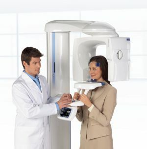 The method is based on the impact of X-rays, and based on the laws of physics, is made "cast" jaw and teeth. And this is due to the fact that literally all the tissues of the body, absolutely differently refract or miss X-rays.
The beam itself, when it is passed through the patient, is caught from the back by a special fixing device.
Depending on the frequency of the wave, some areas darken, while others, through which the rays could not easily pass, in particular the bones, remain bright.
With a series of pictures taken by the computer, after a certain time, a special model is constructed in three-dimensional space, instead of two-dimensional.
The results of the study can be stored both in the patient on the disk and in the clinic.
These are very important data that can greatly facilitate the life of the patient and the treating specialist.
So, for example, having come to the otolaryngologist, you will already have a picture of the maxillary sinuses.
Tomography of the jaw differs from the general, in that here you do not need to lie in a large apparatus and wait while X-rays are spinning around you.
All that is needed is simply to go to a special stand where a 3-d model of your jaw will be shot with the help of a plate, after which it will go to the computer of the doctor, and you together with it will be able to decide what is or should not be done withthis further.
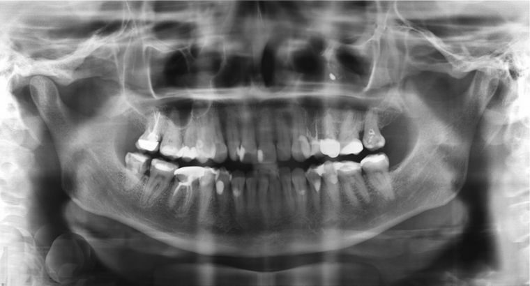
In the photo 3d image of the teeth
How the CT / 3D tomography of the teeth is performed:
The variants of the study
3d tomography of the teeth can be performed on the basis of two different techniques:
- The spiral computer method .This species is very simple to explain by its name. The tomograph simply moves in a spiral, as if examining the head from all sides, and simultaneously emitting X-rays to a special surface where there are special sensors. The doctor from the cabin controls the speed of rotation, with an increase in which increases and the studied surface area. Thus, it is possible to significantly reduce the radiation radiation to which the patient is exposed, and greatly accelerate the very process of scanning.
- Study based on the multi-layered is some improvement on the previous version, with the addition of new features. This time, the sensors are not located on the entire surface, but are clearly arranged in a row, so that it is possible to examine each "layer" of the body and bone tissue. Such a tomograph is able to scan up to 200 completely different layers. This allows you to monitor all the processes that occur in the patient's body in real time, literally looking at the monitor. And thanks to this improved technology, for a couple of turns you can explore the whole body. This same technology allows you to observe the arrhythmia "from within".
Contraindications and indications for the use of the
procedure The procedure is strictly contraindicated:
- to pregnant women, since radiation can damage an undeveloped fetus;
- to people who have a strong allergic reaction to iodine, which is used for tomography.
Computer tomography is assigned:
- to those who suddenly began to show neurological symptoms, especially if it has an avalanche;
- to all those who have blood circulation in the brain, also a passing one;
- when received by the patient in a recent perspective, any in severity of brain trauma;
- to all those who have high blood pressure inside the skull;
- if the patient has epileptic seizures, or any other abnormalities in this path;
- to those who have major brain problems;
- for any symptoms of brain damage, and or, its departments;
- in case of suspicion of the patient for cancer, as well as other diseases that can only be detected by x-ray;
- if the patient suspects any anomalies of cranio-vertebral parishes;
- if the patient heals, or has recovered, from any strong and dangerous diseases of the brain.
The word to the patients
From the practice of patients who have been taken 3d teeth shots.
I would like to tell you about how my affairs were during the operation, which the doctors called "resection of the tooth root."I went to the clinic about this, but I was very much afraid that doctors mistakenly can remove a healthy tooth when it can still be cured. However, after going to tomography, I was shown that it is unlikely to cure him.
Unlike what I expected, the tomography looks completely different. Instead of a gigantic device, I saw a small disc, which could not but rejoice, because thanks to our media, you really start to be afraid of what is not, in this case, irradiation.
Did everything quickly enough, and the doctors with the help of the received 3-d model of the jaw were able to determine that prosthetics is possible. In general, it was not scary!
Alina Sergeevna
I visited several clinics, since I had to do a few prosthetic prostheses, and I did not like everything at all. Finding myself, it seemed to me the ideal option, I was sent to a tomography.
At first I was terribly afraid, it seemed that either it would be very painful or dangerous, because all I knew about the tomography machine was that it emits X-rays.
And then, this is from the physics lessons at school, I myself have not encountered it in my life. And you know, I was very surprised. A small plate, to which they asked to press their teeth, though it was a little painful, but not at all what I expected! Fears did not come true.
Klimansky AS
Advantages and disadvantages of
Computer tomography of the tooth allows you to get the best picture of teeth and jaws, without distortion. And the patient for his part, receives the smallest dose of radiation, from all possible. This allows you to make it to children, and adults without exception.
Pros of tomography: 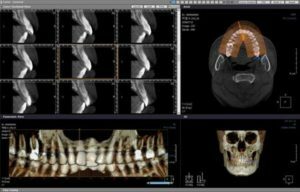
- allows to make sure of absence of deep violations of the dentition;
- identify teeth in the mouth that have not yet erupted;
- see any tumors, dangerous and not very;
- clearly indicate that it was damaged in case of injury.
Among the shortcomings can be designated:
- high cost, especially in private clinics;
- possible human factor.
Price issue
Computer tomography prices depending on the nature of the procedure:
- orthopantomogram( without other procedures) will cost from 1050;
- tomography on a computer( with 2 jaws) will cost from 3300;
- computed tomography on one jaw will cost from 2200;
- computed tomography, which includes only a couple of teeth, will cost from 1100;
- cost of TWG starts from 1550;
- record a picture on any media, it will cost from 500.
All prices are in rubles.
