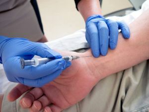
 Phlebography is an study of the vascular system using fluoroscopy .
Since the standard X-ray does not have the ability to be displayed on the resulting picture of the structure of the vascular system, the following method was invented: before the X-ray, the patient is first injected with an X-ray contrast substance into the vein of the legs, which allows the vein to be isolated in the image.
Content of the
- procedure
- content
- testimony Indications for the
- study When it is not possible to use the phlebography
- Preparing the patient for the phlebography
- Procedure procedure
- Patient feedback
- Procedure price
- Conclusions
The essence of the
procedure The presence of contrast in the veins allows the doctor to see the presence of defects in the veins.
Radiopaque phlebography allows to diagnose:
- deep vein thrombosis;
- presence of an embolus;
- various congenital and acquired vascular anomalies;
- check the functionality of the venous valves, analyze their patency, lumen and nature of filling.
You can also reflect the complete picture of the number of communication veins. It makes sense to perform a procedure for visualizing the vein before the bypass.
Phlebography is often performed by for questionable results of non-invasive research methods of .
It should be noted that phlebography is not a screening test due to invasiveness and the presence of X-rays. Nevertheless, it is possible to use a large-frame fluorograph, this will reduce the radiation impact on the patient and the doctor making the shot.
Thus, the duration of phlebography by this method can reach 60-70 seconds .During this time it is possible to make up to 9 pictures.
It is performed in a slightly different way, with the help of special sensors fixed in the sternocleidomastoid muscle, in conjunction with the cardiogram and 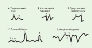 , other studies of the heart.
A record of the curves( phlebogram) is kept, which shows possible violations of the flow of blood to the right atrium and outflow to the ventricle.
Analysis of these curves provides an opportunity to identify many vascular diseases.
Indications for the
study. Phlebography is prescribed with:
- varicose veins with unconvincing functional tests;
- the need for intravenous drug management for sclerotherapy;
- for recurrences of varicose veins;
- in postthrombophlebitic disease.
The procedure can also be performed if a patient has an elephantiasis syndrome to analyze the structure of his venous system.
 About how effective anatomical pillow Varifort from varicose veins you can learn from our article.
 How not to turn into a medicine medicine Warfarin - instructions for use, contraindications, possible complications and other useful information about this drug.
When phlebography of
can not be used With the development of effective non-invasive methods of research, phlebography gradually took second place. In connection with the presence of many contraindications and side effects, it will be used only in extreme cases.
In the future, a more secure method for phlebography is expected to be developed.
The method of investigation has a number of contraindications:
- allergic to contrast agent;
- hypersensitivity to iodine and iodide preparations;
- exacerbation of kidney or liver disease;
- inflammatory processes in the zone of trophic disorders.
Relative contraindication is the advanced age of a person, pregnancy, the presence in the history of allergic reactions of different genesis, atherosclerosis of the vessels, chronic diseases of the kidneys or liver.
 Occurrence of of thrombophlebitis, abscess, skin necrosis, phlegmon is rare. With insufficiently qualified procedure, tissue damage is possible. Sometimes there is an infectious infection, inflammation of the veins, the formation of blood clots and kidney damage.
Allergic reactions are often observed, so the doctor is always ready to identify the symptoms of anaphylaxis( a sharp fall in blood pressure, a breach of breath, collapse, loss of consciousness).
It should be remembered that in patients with diabetes, in particular, those who take metformin have an increased likelihood of complications.
Preparing the patient for phlebography
Before the procedure, the patient should not eat anything for four hours, and drink only water. The presence of an allergy to iodine is checked. If the patient agitates excessively, he is prescribed a sedative.
Before the procedure, the patient or his relatives must necessarily give written consent for phlebography, as well as conduct the patient's analysis to determine the indicators of blood clotting.
A few minutes before the procedure the patient should go to the toilet( urinate), undress and put on a robe.
During the procedure, some painful sensations are possible, to reduce which the patient is prescribed analgesics.
Procedure of
The patient is placed horizontally on the back, there should be no support under the legs. Limbs should be relaxed and not move.
For a better introduction into the vein of contrast, in the area of the ankle a tourniquet is applied.
On the back of the foot, physiological saline is injected into the superficial vein. Then, slowly, for 90 seconds or more, a contrast is introduced.
If it is not possible to perform venipuncture , carry out the venesection. It is necessary to warn a person about the possible occurrence of nausea, burning and other symptoms during or after the introduction of contrast. In this case, the patient should immediately inform them about the doctor.
With the help of fluoroscopy, the doctor observes the spread of the coloring substance, and takes the necessary pictures. After that, the limb is quickly lifted and  is injected with a solution to remove the contrast agent.
The needle is removed only when fluoroscopy shows that there is no contrast in the leg venous system. Then apply a bandage to the injection site.
Soon the patient can return to the usual way of life, provided there are no complications and presence of deep vein thrombosis.
Patient Reviews
What patients say that have gone through the phlebography procedure.
It was necessary to find a suitable vein for shunting. The doctor said that it would be necessary to make a phlebography. I did not particularly understand the essence of the methodology and immediately agreed to the procedure.
But as it turned out, everything was not so simple. In addition to a number of preparatory actions and a very complicated process of phlebography, I was still tormented by mild nausea, weakness and depressed state for a whole day.
As it turned out, it often happens, if you do phlebography to people of my age. Nevertheless, the shunting was successful and soon I will be at home.
Vera, 51
I had a suspicion of deep vein thrombosis. I performed ultrasound. But it did not give a clear answer. Then the doctor appointed me a phlebography for an accurate diagnosis. There was nowhere to go, and I agreed.
During the procedure, the doctor found defects in filling the veins, I filled in some deep veins, and there was a non-standard movement of blood through the collateral vessels. Subsequently, the doctor accurately diagnosed - DVT.
Svetlana, 33
Price of procedure
Cost of radiologically contrasting ascending phlebography in Moscow clinics varies from 8000 to 18000 rubles.
Conclusions
 The phlebography of the lower and upper is one of the best methods for studying the of the human venous system. Practically in any case by means of the given procedure it is possible to put the exact diagnosis.
The only drawback of phlebography is some side effects and the likelihood of the presence of factors that change the results of the study. Some factors depend on both the doctor performing the procedure and directly from the patient.
Possible tight embedding of the bundle, delay in taking pictures, insufficient introduction of coloring matter and even accidental movements of the patient.
This indicates that if you follow strictly instructions, then undesirable moments will be minimized, and you will go through the phlebography effectively and without side effects.
