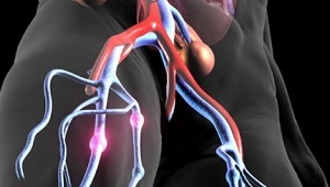
 Venous thromboembolism is considered one of the most dangerous vascular diseases. The pathological process of this disease consists of two directions: deep vein thrombosis and pulmonary artery thromboembolism.
Deep vein thrombosis is the result of a blood clot in the deep veins of the legs and pelvis. The formed thrombus prevents the flow of blood through the veins. As a result, there is a risk of formation of new clots, which can cause serious deterioration.
Most often, the deep veins of the lower limbs and hands are thrombosed, then in the sequence of the decrease are thrombosed:
- cerebral sinuses;
- veins of the abdominal cavity;
- veins of the small pelvis.
Contents
- Reasons and pathogenesis of
- What is venous thrombosis and PE?
- Symptoms depending on location
- Diagnostic criteria and methods
- Medical assistance
- What is the danger and what are the consequences?
- Preventive methods
Causes and pathogenesis of
The cause of blood clots is an imbalance between systemic anti and procoagulants. Also, venous thromboembolism appears as a result of an imbalance between antifibrinolytic and profibrinolytic activity. These imbalances are caused by the following pathogenic factors:
- venous blood flow disorder;
- change in blood composition;
- damage to the vascular wall.
This disease is based on the merger of acquired and congenital risk factors. Congenital factors include 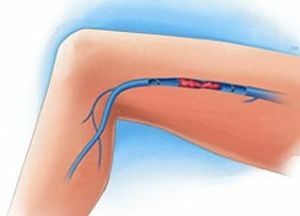 various gene mutations and a shortage of antithrombin and protein. With regard to acquired risk factors, they include previously transferred surgical operations, injuries, obesity, pregnancy and nephrotic syndrome.
Very often thrombi occur in people who smoke, because they have a violation of the tone, narrowing of blood vessels and increased blood viscosity. Also in a large risk area are people with oncological and cardiovascular diseases( coronary heart disease, hypertension, atherosclerosis).
What is venous thrombosis and pulmonary embolism?
Venous thrombosis is an acute disease caused by blood clotting with the subsequent formation of thrombi. The causes of deep vein thrombosis are such factors:
- overweight;
- advanced age;
- smoking;
- carried by cesarean section;
- use of drugs that affect blood clotting.
Favorable conditions for the development of thrombosis is a prolonged bed rest. In this case, the blood flow slows down and a reverse flow of blood to the heart occurs.
PE( pulmonary embolism) is a clotting of the pulmonary arteries by thrombi. The formation of these thrombi often occurs in large veins of the pelvis or lower limbs. Three factors contribute to the appearance of this disease:
- inhibition of fibrinolysis;
- blood flow disorder;
- dissociation of the vascular wall endothelium.
Symptomatic versus localization
Venous thromboembolism of the lower limbs is accompanied by edema, increased skin temperature and swelling of the 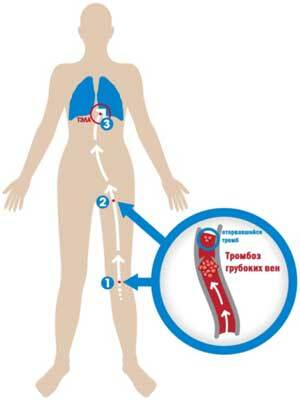 veins with a change in skin color. Most often, the symptoms of this disease are localized around the place of edema and redness. But sometimes the general symptoms can also be manifested:
- sensation of heaviness in the limbs;
- shortness of breath and shortness of breath while walking;
- cough( if there is PE).
Symptoms of venous thromboembolism depend on its location:
Thrombosis of the inferior vena cava .Symptoms are manifested in the form of bypass venous circulation and edema of the lower limbs. In case of thrombosis spread to the hepatic veins, jaundice and ascites appear.Diagnostic criteria and methods
Venous thromboembolism is diagnosed by several methods:
- of deep vein ultrasound with compression of .This diagnostic method has a sensitivity of 95%.Therefore, the results will depend on the qualifications of the specialist who conduct the diagnosis. In the presence of thrombosis, it is impossible to squeeze the vein with an ultrasound transducer. Difficulties in diagnosis can occur with repeated blockage in the deep veins of the lower extremities.
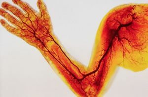
- Phlebography is the most accurate method for diagnosing thromboembolism of the lower extremities. The diagnosis is made in the presence of filling defects in the lumen of the vessel.
- Venezography .This is the most accurate method of diagnosis, but it is used infrequently, as it is an invasive and even painful method. With this method, there is a risk of allergic reactions.
- Duplex scanning .It includes two research methods: Doppler blood flow measurement and ultrasound examination of blood vessels. A significant disadvantage of this method is that it has very low sensitivity in the study of asymptomatic patients. The use of this technique is contraindicated in the presence of fixing tires and bandages on the lower limbs.
Sometimes for screening, other screening methods are used in the form of impedance plethysmography and scintigraphy. The latter method is used with labeled proteins that are capable of binding to a glycoprotein.
Medical assistance
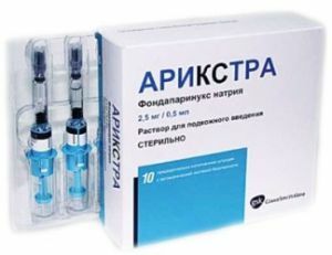 To date, 90% of patients with venous thromboembolic treatment are treated with anticoagulants.
Other treatments are used for individual patients in a particular condition. Such patients are people who have contraindications to therapy with anticoagulants.
In general, the following methods are used to treat VTE:
- Initial anticoagulation .It is carried out for five days with the appointment of Fondaparinux and Heparin. After five days, early maintenance therapy is used. In this case, antagonists of vitamin K are prescribed. To achieve a certain therapeutic effect, the drugs should be consumed for five to seven days. After this, the initial anticoagulation is stopped.
- Systemic thrombolysis .This method of treatment is more often used for PE because it reduces the volume of the clot more quickly. Its essence lies in the dissolution of thrombi under the action of a certain enzyme, which is introduced into the blood. The disadvantage of this method is the risk of hemorrhagic complications. Systemic thrombolysis is performed by intravenous administration.
- Catheter Interventions .There are three types of catheters that have the effect of thrombolysis: transfemoral, trasbrachial and triacarial. This method of treatment is very expensive, as it requires complex outpatient supply, equipment and qualified personnel.
What is the danger and what are the consequences?
After a venous thromboembolism, the vascular patency is restored, but the venous function may be weakened.
This is due to the fact that the venous valves were previously destroyed. As a result, secondary varicose may appear, which will lead to an increase in venous hypertension. If the treatment is not performed in time, then there is a risk of manifestation of postthromboembolic syndrome.
Patients who have suffered pulmonary thromboembolism may develop complications such as prolonged thromboembolic pulmonary hypertension. This deterioration is a type of high blood pressure in the vessels of the lungs.
Preventative methods
Prevention of venous thromboembolism is to prevent the risk of its occurrence and to determine the risk category( low, moderate, high) in the presence of the disease. 
One of the methods of prevention is elastic compression of the lower limbs. In this case, you can apply elastic compression stockings and knee socks. They help to normalize the venous outflow and distribute the pressure along the length of the entire lower limb. You can also use special medical jersey.
Another method of prevention is intermittent pneumatic compression, which is performed using a special compressor.
Inflating the cameras is a very useful effect, especially if there is no muscle contraction in the limbs for a long time. This method helps to increase the rate of muscle blood flow, even in immobilized patients.
