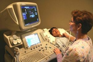
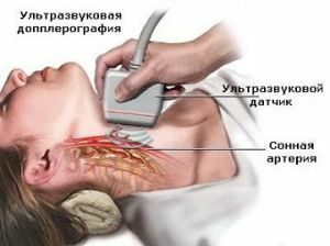
 Duplex ultrasound scanning is an combination of Doppler ultrasound with conventional ultrasound scanning of .
This type of study allows the doctor to assess the condition of blood vessels, see the movement of blood in the vessels and determine the speed of blood flow, measure the diameter of the vessels and detect the presence of blood flow disturbance due to blockage of the vessel.
Traditional ultrasound scanning is based on the use of ultrasonic waves, inaudible to the person, reflected from blood vessels.
The computer captures these waves and converts them into a two-dimensional image.
In the Doppler ultrasound study, the determines the rate at which sound waves are reflected from the moving blood elements of the .
The ultrasonic waves are stressed on the blood elements, for example, erythrocytes, and their reflection, and the speed of the ultrasonic wave depends on the velocity of the blood elements.
Based on this type of study, a two-dimensional image is displayed showing the presence of obstructions to blood flow, such as narrowing the lumen of the vessel with an atherosclerotic plaque.
The image formed on the basis of a duplex examination can be colored, indicating to the doctor the places in which the blood flow is disturbed, its speed, and also the direction.
This type of ultrasound scan uses for diagnosis:
- of occlusive carotid artery diseases( cerebrovascular disorders);
- of various diseases of the vessels of the lower limbs( obliterating atherosclerosis, arterial aneurysms, deep vein thrombosis);
- of upper limb vessels diseases( Raynaud syndrome, upper chest aperture syndrome, vein thrombosis);
- of occlusive diseases of the aorta and iliac vessels, varicose veins of the legs, aneurysms of various parts of the aorta.
 When troxevasin ointment is unrivaled. Instructions for use, pros and cons of medication and other information in our article.
 This should be known if you have been prescribed Gepatrombin G - instructions for use, reviews of doctors and patients, an analysis of the pros and cons of the drug.
Contents
- Study of brachiocephalic arteries
- Investigation of lower limb veins
- Duplex scanning of thyroid vessels
- Scanning of abdominal vessels
- Duplex scanning of small pelvic vessels
- Study of renal vessels
- Indications and contraindications for procedure
- Preparation for duplex scanning
- Procedure procedure
- Cost of procedure
Study of brachiocephalic arteries
Duplex scanning of brachiocephalic arterium is one of the main methods of ultrasound diagnosis of blood vessels that provide blood supply to the brain.
Duplex scanning of cerebral vessels also allows assessing the condition of general, external and internal carotid and vertebral arteries in the neck.
This method is a combination of visualization of blood vessels to surrounding tissues and studies of hemodynamic parameters using color  Doppler coding.
Using duplex scanning of brachiocephalic vessels, blood distribution in the lumen of vessels of any caliber can be estimated.
Including large arteries, peripheral vessels, subcutaneous capillaries.
Study of lower limb veins
Duplex scanning of the veins of the lower limbs is indispensable for modern medical diagnosis method.
This type of study is conducted primarily for the of the determination of the condition of the lower extremities veins and the functional consistency of the blood flow. This method is widely used in phlebology and vascular surgery to identify the pathology of the veins of the lower limbs.
Most often the procedure is performed in the presence of varicose veins. With duplex scanning of the veins of the lower limbs, the doctor evaluates the degree of permeability of the veins, the functional consistency of the valvular veins apparatus, and the speed of blood circulation.
This study allows you to accurately find out the cause of of a particular pathology of the vessels of the lower extremities and to assign the right type of treatment with maximum efficiency.
Duplex scanning of thyroid vessels
Duplex scanning with ultrasound examination of the thyroid gland allows the doctor to assess the blood supply of the organ and determine the presence of such conditions as chronic thyroiditis and goiter.
Investigation of nodal formations with the help of duplex scanning allows detecting malignant processes.
Scanning of abdominal vessels of the abdomen
With duplex examination of the abdominal cavity, tumor formations can be detected. Sometimes this technique is decisive in evaluating ultrasound data.
Duplex scanning allows revealing exfoliating aneurysms and atherosclerotic plaques when examining the abdominal aorta.
With ultrasound scanning of the liver, the duplex makes it possible to examine the vessels of the bile duct system and evaluate the direction of blood flow in the portal vein, as well as the condition of the porto-caval anastomoses if the patient has cirrhosis of the liver or portal hypertension.
Duplex scanning of small pelvic vessels
 It is quite widely used in clinical practice to use a duplex study of prostatic vessels in prostatitis, with suspicions of a gland abscess, with prostate adenoma.
Men also have a study of the plexus of the plexus in the prostate. Duplex scanning is used to diagnose various ovarian cysts, assess their normal function and supply blood to various pathological lesions in the walls and uterine cavity.
Study of Kidney Vessels
Duplex study is indispensable in the search for renal veins and arteries, as well as in the study of intrarenal vessels. Often, the goal of duplex scanning is to determine the normal functioning of the kidney and its viability in general.
Indications and contraindications for the procedure
Duplex study of the vessels of the lower extremities is carried out:
- in the presence of signs of impaired blood supply to the limb, its ischemia: sensation of chilliness, distortion of sensitivity, numbness;
- for traumatic injuries of the vessels of the limbs;
- in the presence of signs of vascular aneurysms;
- for pain arising during walking;
- with hereditary predisposition to thrombosis;
- when changing the color of the limbs.
Duplex vascular scanning, unlike most other instrumentation diagnostic methods, does not have any contraindications and age restrictions.
In this regard, one patient within one day can be examined as many times as required for an accurate diagnosis. Duplex scanning is absolutely safe for both adults and children.
Preparing for duplex scanning
Preparation is necessary only with duplex examination of the vessels of the abdominal cavity, since in this case it is necessary to prepare the intestine, this is due to the difficulty of passing ultrasound through the contents of the intestine.
In all other cases, there is no need for special preparation for the procedure.
Procedure

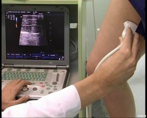
On the photo, duplex scanning of the lower extremity veins
The patient is laid on the couch in a convenient position for him, while the head is in a slightly elevated position. The doctor performing the procedure lubricates the sensor of the device with a special gel to facilitate movement on the patient's skin and improve the transmission.
The information received by the sensor is converted into a digital signal and generates an image on the monitor screen. Often during the procedure, a special sound is heard, characterizing the state of blood flow in the vessels.
The whole procedure usually takes half an hour, but the time can vary depending on the size of the scanned section.
The cost of the procedure
Duplex scanning can be done in many medical institutions, the price of research regarding is democratic:
- Medical Center in Marino - 2100 rubles.
- Kuntsevo Center under the direction of DI Dikul - 2100 rubles.
- Miracle Doctor at School 49 - 1980 rubles.
- Clinic number 1 in Khimki - 1840 rubles.
- Clinic "АВС Медицина" on Leo Tolstoy - 3080 rubles.
- Treatment and Diagnostic Center "NovoMed" - 1900 rubles.
