Endometriosis is one of the most frequent and mysterious diseases in gynecology. A large number of women, having all the symptoms of a disease, do not know that pain, copious periods, infertility and many other things are connected with this pathology. The most informative diagnosis of the disease is laparoscopy and some endoscopic methods of investigation. But all these are operational interventions, and routine methods can not be routine for a number of reasons. Ultrasound examination( ultrasound) of the pelvic organs is widely used in gynecology. This is a simple, inexpensive way that takes about 15 - 20 minutes of time for a woman and a doctor, does not imply special preparation. But it is important to know when to do ultrasound in endometriosis, if there is a suspicion of this pathology, in order to confirm or deny the diagnosis as accurately as possible.
For what purposeprescribe
Symptoms of all gynecological diseases are similar in many respects, and only certain accentuations or signs can lead a doctor to think about a particular pathology.
Why is ultrasound
Endometriosis brings a lot of inconvenience and problems to a woman. Treatment of this pathology is complex, long and multicomponent. Therefore, in order to accurately verify the presence of endometriosis, use a variety of methods, including surgical interventions. And only after the histological confirmation of the diagnosis( sometimes in a vivid clinical picture) can treatment be prescribed, in most cases, hormonal.
Ultrasound examination of pelvic organs with a high probability may indicate this pathology, without resorting to more invasive and life-threatening methods for women. In order to understand the ultrasound for endometriosis on which day it is best to do, it is important to know the mechanisms of endometrial transformation and to assume the places of other foci.

Detection of pathology
The cause of the development of endometriosis is not fully understood. But, based on those data that are known to medicine, the diagnosis, treatment and prevention of pathology are under construction.
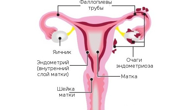
It is believed that the sites, rather even individual cells, the endometrium - the inner layer of the uterus, for some reason, start in different ways into other parts of the organs of the reproductive system of women. It happens retrograde( through the fallopian tubes), with blood, lymph flow and other means. As a result, where normal endometrium should not be, it appears and begins to function, changing under the influence of hormones just as in the uterine cavity.
Normally, the inner layer of the uterus is rejected monthly with the appearance of a small bleeding, then it is restored. And so cycle after cycle. Migratory sites behave the same way. For example, if the ectopia touches the ovaries, then, as a result of such a secretory process, a cyst is formed, filled with dark brown, usually stiff, contents. It grows over time - from a size of less than a millimeter to tens of centimeters.
The pelvic ultrasound for the detection of endometriosis is performed for the purpose of differential diagnosis in the following situations:
- in painful menstruation;
- with abundant and prolonged discharge during critical days;
- if present for several days before and after the monthly brown smear;
- for infertility;
- if there is discomfort, sometimes even pain during physical exertion, sexual intercourse, etc.
Ectopic patches can be localized theoretically on any organ or structure in a woman. Sometimes the foci are found on the baking, often - on the loops of the intestine and on the inner lining of the abdomen( peritoneum).As for the female genital area, the affected areas can be found in the following departments:
- external genitalia and perineum,
- vaginal mucosa,
- cervix and cervical canal,
- muscular part of the uterus( this type is called adenomyosis),
- tubes,
- ovaries.
To search for other affected areas of
, the endometriosis of each part is characterized by certain signs on the ultrasound. Having discovered the pockets in a particular place, the doctor determines the further tactics of treatment.
When localization of foci in the area of external genitalia and perineal ultrasound is performed to determine the degree of prevalence of the process. Exactly the same goals are pursued in the detection of endometriosis inclusions on the mucous membrane of the vagina.
Often, especially after childbirth and surgery( scraping the uterine cavity, abortion, etc.), foci can be identified on the cervix. For their further investigation, colposcopy is performed, but to determine if the process is deep down into the cervical canal, ultrasound of the small pelvis is performed using a vaginal sensor.
During a gynecological examination, an increase in the uterus body can be diagnosed in women. This may indicate pregnancy, fibroids, occurs in the mnogogorozhavshih and after caesarean section, as well as in the case of adenomyosis. At the same time, the uterus body acquires a globular shape - one of the most significant and frequently occurring diagnostic signs of endometriosis, including the results of a pelvic ultrasound.
Localization of foci on the ovaries eventually leads to their proliferation and the formation of cysts. Sometimes they reach a size of more than 10 cm. Ultrasound with endomeriosis, on which day of the cycle would not have been performed, gives high reliability precisely at this location. Often in these cases, both ovaries are affected, simultaneously or with some interval. Ultrasound examination of the pelvis with a confidence in 80 - 90% makes it possible to distinguish endometriotic cysts from other neoplasms.
When to do
In order to identify the pathology with the greatest probability, it is important to determine the most appropriate time for the study. For this, it is necessary to understand how endometrial transformations occur during the menstrual cycle. After all, in fact, endometriosis - small scattered its foci.
At the beginning of the cycle, the upper layers of the endometrium are rejected. During this period, its thickness is minimal, usually a millimeter or less, in such cases it is said to be linear. With each passing day, the endometrium grows, and by the end of the cycle it reaches a maximum value of 15-25 mm on average.
If the ultrasound at the beginning of the
cycle Classically, an ultrasound examination of the pelvic organs in women is classically considered to be informative for 5-7 days. Identify endometriosis at this time, too, you can, but this applies only to its special localizations and common forms. For example, with the location of foci on the ovaries, as well as with pronounced adenomyosis. Also performing a study at the beginning of the cycle, the doctor can presume endometriosis by indirect signs, for example, in the presence of endometrial hyperplasia, globular shape of the uterus, and others.
 We recommend reading an article about ultrasound during the period. From it you will learn about the optimal days for the examination, the indications for the procedure, the reasons why ultrasound can not be postponed.
If ultrasound at the end of the
cycle The most reliable information and a vivid picture of the pathology can be obtained by performing ultrasound on the eve of the month, for 23 - 25 days. At this time, endometriosis will be largest, its symptoms will be as pronounced as possible.
On what day it is better to do ultrasound in endometriosis, the attending physician should decide on the basis of the clinical picture of the complaints and the alleged diagnosis. This is the only way to confirm or deny pathology with a high degree of probability, without resorting to other methods of research, and also to establish, if necessary, the prevalence of the disease.

