 Caries refers to dental diseases, a characteristic feature of which is the destruction of hard tooth tissues. In the initial stage, the pathological process is limited to the demineralization of the enamel.
Untimely access to the dentist leads to the destruction of dentin and the formation of a carious cavity, later the inflammatory process develops in the pulp and periodontium.
If the carious area is located in the proximal zones, it can not be determined independently. Therefore, the superficial caries gradually changes to more advanced stages of the disease, the final stage of which may require a depulpation procedure.
In modern dental institutions there are special diagnostic methods for detecting the earliest stages of caries with initial damage to the enamel, which allows preserving the integrity of the dentition, provided that timely preventive examinations are carried out.
content
- general approach
- Visual inspection
- Percussion
- Diagnosis using the probe
- Drying
- Staining
- tooth surfaces Application roentgen
- Test for pain by changing the temperature
- Test low
- discharge current Translyuminestsentsiya
- autopsy patient tooth
- Differential studies
- Difficulties in the wayto the statement of the correct diagnosis
General approach of
Diagnostics consists in the collection of anamnesis and the examinationa patient's use of various techniques that help define 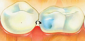 presence and degree of development of caries.
Survey of a patient includes questions about the nature and timing of the onset of pain, the presence of chronic diseases, the number of affected teeth, previous treatment and its effectiveness.
After preliminary familiarization with the situation for the final diagnosis, the patient is examined using the appropriate research methods. Their choice depends on the degree of carious lesion and the localization of cavities.
There are four degrees of destruction of hard tooth tissues:
- The initial stage of is characterized by the appearance of a white spot;
- surface lesion changes the integrity of the enamel, visually reveals a slight defect in the tissue;
- average caries destroys dentin, the patient complains of the appearance of pain when taking sweet, salty, hot or cold food;
- The deep destructive process of is characterized by the presence of a large carious cavity, while the pulp is not affected.
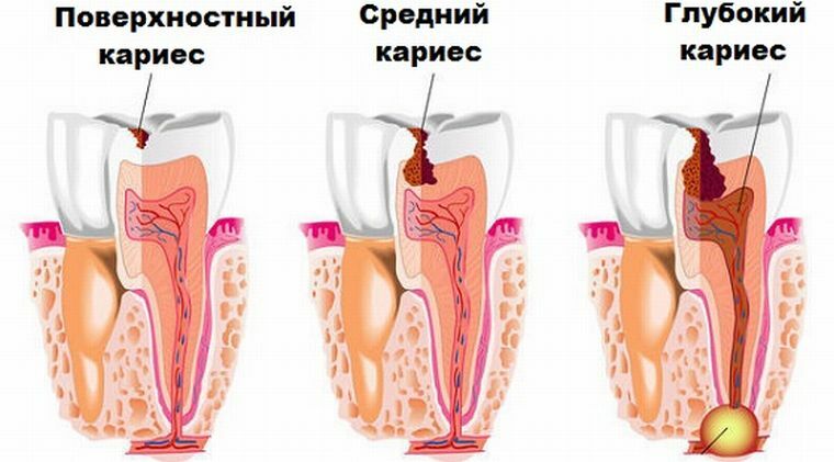
Recognition of medium and deep caries can be visualized with the help of mirrors and sounding, but the initial and surface stages are practically asymptomatic; therefore, in addition to the above methods,
- drying is usually used;
- transluminescence;
- percussion;
- vital staining;
- thermal probe;
- radiography.
Visual inspection of
Most dentists agree on the idea that the teeth should be treated after a preliminary stone removal session. This is due to the difficulty in establishing a correct diagnosis when the surface is covered with a touch of plaque or tartar.
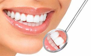 Visual inspection is performed using a special dental mirror, which reflects light rays on darkened areas of the oral cavity. With this tool, distal surfaces can be examined in detail.
When the caries appears, the enamel changes color. At the initial stage, characteristic white spots are formed. The structure of the surface loses its properties, becomes dull and rough. Later, brown and black dots or furrows appear on the teeth. If the changes occur inside the tooth, you can see a darkening in the place where the carious cavity is formed.
A single examination is usually not enough, as some diseases can be masked for caries, for example, pulpitis, in order to avoid an error, differential diagnostics is assigned with the help of certain test methods as the case may be.
Percussion
In medical practice, this term means tapping certain areas. By the nature of the sound can determine the presence of damage in the structure of the tooth.
Diagnostics using the
probe The use of the probe allows differentiating caries and pulpitis, as in the second case there is a sharp pain when moving in the 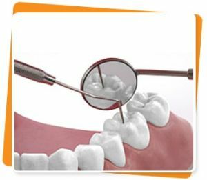 in a certain direction.
In caries, sensitivity is manifested when examining the walls and cavities of the enamel-dentine zone at the site of the focus. Also this device helps to determine the nature and depth of the carious process.
The density of the affected walls indicates the chronic course of the disease, while the softening of the tissues indicates the activation of the destructive process.
Drying of
This method is effective only in the detection of the superficial form of the disease. From the tooth to be examined, plaque is removed, further measures are taken to prevent saliva from entering it, after which the object is exposed to air currents.
During the procedure, all excess water evaporates, and the caries stain acquires a matte shade. The affected surface becomes slightly rough.
Teeth surface coloring
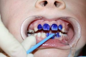 For the purpose of determining the depth of microdefects of enamel, the teeth are treated with a 2% solution of methylene blue. Damaged places are painted in different shades of blue, the intensity of which depends on the degree of damage. This procedure is carried out after preliminary cleaning of the survey site.
With this method, it is possible to determine the presence of changes in the dentin at the bottom of carious cavities. For this, a tampon moistened in a 2% solution of methylene blue or 1% solution of potassium iodide is placed in the depth of the formation.
And after a couple of minutes evaluate the result. With the same purpose, indicators based on magenta are used. Only the painted damaged surface in this case acquires a pink tinge.
X-ray application
This diagnostic method is ideal for detecting deep caries. On the x-ray image you can see the depth of the tooth lesion and 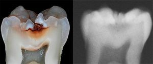 the distance from the hearth to the pulp.
The method is well proven in the diagnosis of latent caries in those places where visual inspection does not give a proper result, for example, between the teeth, under the crowns, near the neck of the tooth.
Changes in the tooth are revealed by the characteristic clearing in the cavity of hard tissues. But if the focus of the disease is not caries-forming, then it will not be displayed on the x-ray.
Sore test for changing temperature conditions
With periodic discomfort in one part of the dentition, the surface is alternately treated with cold and hot water.
If there are unpleasant sensations, caries is diagnosed, when the pain is long and acute, this indicates the appearance of pulpitis. Despite a number of advantages and simplicity, this method is rarely used in modern dentistry.
Low-current test
Electrodontometry is sometimes used. It is used to determine the degree of involvement of pulp in the carious process. The method consists in the action of a small discharge of current on the affected area.
Depending on what kind of voltage is required for the appearance of a reaction from the tooth and the tingling response, you can determine the normal condition, inflammatory process or necrosis of the pulp tissue.
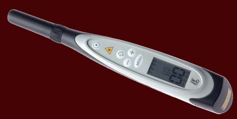
DIAGNOdent pen - apparatus for the diagnosis of hidden caries in the fissure area
Transluminescence
This method helps to determine the presence of destructive processes and the general state of the enamel. There are two types of diagnostics: 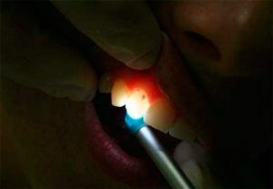
- Fibrooptic examination is conducted in the dark. It consists in enlightening the tooth with a bright cold light beam. Carious sites are distinguished by dark zones in the overall picture of a healthy surface.
- Luminescence of .Under the UV rays passed through the Wood filter, the carious areas are displayed in dark spaces against the snow-white background.
Autopsy of a sick tooth
Fissurotomy is performed only in those cases when the doctor is confident of the presence of a carious cavity. The opening of the fissure area with the help of a special drill helps to assess the extent of the lesion of the site.
If there are only spots on the surface and there is no way to confirm the presence of the disease by other methods, some doctors take a waiting attitude and postpone the procedure until more obvious signs of the disease appear.
Differential research
Differential diagnosis, with the aim of distinguishing caries from other diseases with similar symptoms, is performed using a number of examinations depending on the degree of damage to hard tooth tissues: 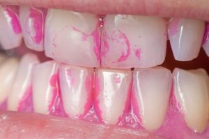
- Staining of the enamel helps to exclude hypoplasia and enamel fluorosis. Penetration of special markers occurs only in the cavity of demineralized enamel, which develops due to the development of the carious process.
- The effect of current and thermo probe are indispensable in determining pulpitis and periodontitis. In the presence of these diseases, the patient's reaction to the manipulation of the doctor will be more pronounced.
- Radiography is designed to detect non-carious lesions of the enamel structure. Also in the picture you can see periodontitis and pulpitis.
Difficulties on the way to the correct diagnosis of
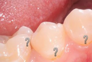 The insidiousness of the disease consists in the fact that superficial, medium and even deep caries can be masked by a seemingly healthy surface wall of the enamel or hiding in approximate surfaces.
However, the most difficult to determine is the initial stage of the disease, a characteristic feature of which is the appearance of white matte spots.
Laser diagnosis of caries:
Usually the medium and deep carious process is noticeable during visual inspection.
Dentists recommend periodically to make independent diagnostics of teeth and the whole oral cavity at home using a conventional mirror.
If there are uncharacteristic brown or black dots or spots on the background of the white tooth row, you should contact a specialist.
It is possible to suspect the appearance of an ail and on the arising uncomfortable sensations with the use of sweet, salty, cold and hot dishes. 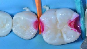
If the pain is short-lived and self-destructive, this indicates the development of caries. In the case of a prolonged manifestation of an unpleasant feeling, one can suspect pulpitis.
And when the pain arises spontaneously, regardless of food intake and passes only after the use of painkillers, you can talk about periodontitis.
Caries is the beginning of destructive processes in the teeth, so it is important to pass preventive examinations on time and to monitor the integrity of the enamel layer.
