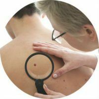
Benign skin tumors or malformations of their development, which are due to a violation of the synthesis and metabolism of melanin, are called moles. They are different: flat, convex or, conversely, smooth, hairy. Birthmarks do not cause discomfort and are considered a cosmetic defect. However, some nevi can easily degenerate from benign to malignant, which threatens human life. Why do birthmarks appear and how to deal with them?
- What causes
- Types
- Red( vascular)
- Dark( not vascular)
- White
- What are dangerous
- Diagnosis
- Removal
- Consequences of removal
- Child
- During pregnancy
- How to remove at home
Why do
- appear Radiation exposure. Normal sunlight can cause skin mutation, as it contains a large amount of ultraviolet radiation, which stimulates the formation of moles. This is especially noticeable in people with fair skin, their body is badly absorbed by ultraviolet, resulting in a mutation of the skin. Many experts believe that the emergence of new birthmarks is associated with exposure to radiation or X-rays, because during life, each person is exposed to radiation every year( for example, fluorography or X-rays).
- Injury of moles. Close or narrow clothes, seams located in uncomfortable places, systematic rubbing with coarse material injures an existing birthmark. It can damage the owner himself - accidentally cut or podkovyrnut. In this case, melanocytes become activated and appear on the surface of the skin with new formations.
- Viruses. Viral infections, such as viral papilloma, play a large role. Its cells contain several genes, because of which they share much more often. The result of growth is unpredictable: it can be either an ordinary wart, or malignant or benign formations.
- Hereditary predisposition. Information embedded in DNA is often the cause of the appearance of hereditary moles. They are the same shape and size as their relatives, even often located in the same places, for example, on the face, neck, folds of hands, fingers.
- Hormonal changes. One of the hormones produced by the pituitary gland strongly affects the release of melanin. So often moles appear in adolescents during puberty, but under the influence of hormones they can independently and disappear from the body. Therefore, during this period of life birthmarks do not carry an oncological danger, this is the usual reaction of the body to the restructuring of the hormonal background.
Kinds of
Birthmarks are congenital - they occur right away at birth, and acquired - are formed throughout life. Most of them appear during pregnancy, adolescence and menopause. Nevi can occur anywhere on the skin, in particular, on the mucous membranes.
Depending on the depth of the layer of skin on which they appeared, they are divided into the following types:
- Borderline - appear on the border of the dermis and epidermis.
- Epidermal - appear on the upper layer of the skin.
- Intradermal - are formed in the deep cutaneous layer( in the dermis).
Nevuses differ in appearance and appearance:
- Flat( melanocytic) - the most common and safe kind of moles. In most cases, these are small, smooth spots of the correct oval shape.
- Organoid( warty) - brown, black or blue, reminiscent of the appearance of warts that stick to the leg and protrude over the skin. With this type of nevus you need to be careful, as it is easy to injure.
- Non-vellucid( convex) - formations of dark color rising above the foot with a flat or rough surface, on which the hair sometimes grows.
Color distinguishes between red, dark and white moles.
to contents ^Red( vascular)
Appear as a result of disturbances in the work of blood vessels - arteries, capillaries, lymph nodes and veins. Depending on which vessel is damaged, neoplasms can be of different colors( from pink to blue-red) and sizes. Capillary birthmarks are superficial and flat, cavernous - bumpy and knotty, embedded in the thickness of the skin.
The most common types of vascular birthmarks:
- Hemangioma( strawberry birthmark) appears in the first month of the child's life in the form of light patches or small swells of red. Over time, the edges of the tumor acquire distinct edges and turn red. Hemangioma usually disappears by 7 years.
- Malformation( stork stings and port stains) are congenital red moles that occur during the first weeks of a newborn's life. Because of vascular malformation, the blood vessels are malfunctioning, which leads to the formation of red color on the skin. The stork's sting arises in the neck, neck, forehead and at the temples of the baby. Its formation is associated with fetal hypoxia, which leads to squeezing of the vessels. Such stains, unlike portworms, disappear on the first year of the child's life.
- Port stains are often located on the hands, face and trunk. They do not disappear with age, but only change their color and become darker in color.
Dark( not vascular)
This kind of moles appears from the excessive production of melanin( the coloring pigment).Formations are plural or single plaques of various colors( gray, blue, brown, etc.) and forms with a keratinized surface.
The following safe species are encountered:
- Lentigo is a common form of pigmented formations, with an equal color from pale brown to brown.
- Mongolian spots - neoplasms are round pigmented spots of a blue hue that are localized in the sacral and lumbar zone. Up to 15 years they independently disappear without treatment.
- Coffee stains are flat small brown moles. The presence of two neoplasms does not belong to pathology, more than 3 - observation and additional diagnostics are required, since they can be a sign of neurofibromatosis( the tumor is formed from nerve cells).
White
This kind of moles appears when the production of melanin decreases. Neoplasms can have different surfaces( rough or smooth) and dimensions. Sometimes light nevi are signs of serious illnesses, but in most cases white moles are an individual feature of human skin that is not dangerous to health.
What are the dangerous
Some types of moles are able to degenerate into malignant formations. These include:
- Blue nevus is a round tight knot with no hair, blue or dark blue, measuring up to 5 millimeters. The location of the birthmark is the face, buttocks and limbs.
- Nevus Ota is a large pigmented entity on the face of a dark brown or blue-gray color. It is localized in various areas of the face( cheeks, cheekbones, upper jaw) and has light interlayers, which creates the effect of dirty skin.
- Dobrail melanosis is a precancerous skin lesion that looks like a single spot of light brown color with uneven outlines. Gradually such birthmark increases and changes color, becomes black or dark brown. It is formed in most cases on the face and other open areas of the body.
- Giant pigment nevus is a formation that has a cracked, bumpy or warty surface from gray to black. Birthmark grows with a man.
- Border pigmentary nevus is a flat dark brown or black hairless knot with a smooth and dry surface with a diameter of no more than 10 millimeters. Basically these dangerous birthmarks are localized on the palms, soles or intimate areas.
- Dysplastic moles - neoplasms of various shapes with a diameter of not more than 1 centimeter. Appear on the chest and buttocks. Transmitted by genetics.
- Papillomatous - a convex mole with an uneven surface and irregular contours. The color varies from light-flesh-colored to dark-brown. Often occurs on the head, on the surface may be hairs.
Diagnosis
The study of the mole allows to determine whether it refers to a borderline, melanocytic species or skin formation has a different etiology.
The most common diagnostic method today is dermatoscopy. The study is performed with the help of a dermatoscope apparatus( a microscope, which increases the surface of the skin several times).During the procedure, the doctor evaluates the following symptoms:
- Size and color of the birthmark.
- Edge structure.
- The presence of asymmetry.
If suspected that the mole is degenerating into a malignant one, a radioisotope study is performed that relates to the non-invasive method of diagnosis. A patient on an empty stomach is allowed to drink a preparation containing disubstituted sodium phosphate, and then, using contact radiometry, the amount of isotope accumulated in the formation and the symmetrical healthy skin region are compared.
There is also a thermometric diagnostic method based on the detection of pathological processes in the skin, accompanied by temperature changes. The difference in temperature of the affected and healthy skin should be 4 degrees.
However, the final decision in the diagnosis of nevi is given to the histological examination, which consists in examining a sample of skin formation obtained after the removal of a mole under a microscope. Thanks to this diagnostic method, the doctor can finally confirm or disprove the malignancy of the nevus.
to table of contents ^Removing
There are several ways to remove moles:
- Cryodestruction.
- Surgery.
- Electrocoagulation.
- Laser evaporation.
- Radio wave emission.
The doctor selects the method taking into account the state of the tumor and its features, takes into account possible risks for the patient's organism as a whole.
Most recently, the main methods of removal were cryodestruction and electrocoagulation. In cryodestruction, the mole is frozen with liquid nitrogen, and electrocoagulation acts on the formation of high-frequency electric current. However, these techniques have side effects: the formation of blisters that appear in healthy parts of the body, both after heat and after a cold.
Surgical removal is performed with normal brown moles, since this method allows to completely excise all tissues of formation from deep layers of skin. Also, surgical removal of moles, similar to cancer for carrying out tissue histology.
Radio wave coagulation is used for more subtle procedures. They remove birthmarks in one motion, and this procedure lasts less than 5 minutes. The advantages of this method is that the patient can go home right after the operation.
Effects of removal of
A few hours after the removal of the birthmark in the wound area, pain can occur due to a violation of the integrity of the structure of the skin. Anti-inflammatory nonsteroid drugs( Ketorol, Nimesulid, Nurofen, Ketanov, etc.) will help to cure pain.
The wound does not require special treatment and care until the joints are removed, which is carried out on the 8-10th day. After this, for the early healing and prevention of scar formation, it is necessary to apply such ointments as Solcoseryl, Levomecol or Metiluracil to the wound.
Until the wound is completely healed, so that inflammation and infection do not occur, it is recommended that the following rules be followed:
- Do not tear or wet the crust formed.
- Do not apply cosmetic products to the wound.
- Cover the wound with adhesive tape or cloth from exposure to sunlight.
Complete healing after surgical removal of the mole occurs during 14-21 days. With the use of other methods of removal, wound healing is faster.
In case of non-compliance with medical recommendations, the wound can become inflamed, which will lead to prolonged healing and scar formation. The signs of infection include:
- Growing pain.
- Redness.
- The exit of pus.
- Divergent edges of the wound.
Sometimes seam divergence occurs, as a result of which the edges of the wound spread and slowly grow together as it is necessary. In this situation, you should consult a doctor who will take the necessary measures.
to the table of contents ^The child
Predisposition to skin formations appears when the baby is still in the womb. The causes of the appearance of nevi on the body of the child are:
- Exposure to the body of a pregnant woman of radiation, toxic substances.
- Hormonal fluctuations in the body of a pregnant woman.
- Predisposition of parents to the birth of moles.
- Urinary tract infection in pregnancy.
On the skin of a child, a red nevus may appear - a hemangioma( vascular birthmark).This kind of neoplasm is not dangerous and will disappear after some time. A special type of skin defect is Setton's nevus, when a white spot( skin without a pigment) appears around the neoplasm. This can be a reaction to the effects of ultraviolet light or the effects of sunburn. Treatment in this case is not required, after several years the spots disappear, and the skin returns its color.
Often the children show convex birthmarks of various colors from light brown to black. Such nevi are easily damaged, especially in places where they can often be traumatized. In this case, it is better to consult a doctor who will remove the birthmark.
- Uneven edges.
- The asymmetry of the nevus.
- Heterogeneous color.
- Large size( over 6 millimeters).
- Quick change in size.
The presence of these signs does not mean that the mole began to degenerate into a malignant, but the expert can only give an accurate diagnosis.
to contents ^During pregnancy
The appearance of a large number of moles in women waiting for a child is associated with hormonal changes in their bodies and is considered the norm.
It is necessary to worry if a lot of birthmarks appeared on the body during pregnancy, which caused inconvenience: itching, flaking, burning, they began to increase in size, change their color, shape, bleed, thicken, become convex. In this case it is necessary to consult a doctor. Perhaps, all the fault is the same hormonal fluctuations, and after birth the mole will again become the same size, but only the specialist can determine it.
to the table of contents ^How to remove at home
Traditional methods of treatment of moles involve the use of various compresses, ointments, infusions, etc. Safe means are those that do not immediately remove the nevus, but eliminate or make it less noticeable gradually. However, in this case, before the treatment, make sure that the mole is not malignant.
If the doctor confirms that the skin education does not threaten health and only carries a cosmetic defect, then you can use one of the following folk remedies:
- Iodine. It is used for sensitive skin. Droplet funds should be applied with a cotton swab to the nevus 2-3 times a day. The procedure should be carried out daily until improvement.
- Garlic. Apply a patch to the nevus with a hole cut out for it. Then squeeze the clove of garlic with a press, wipe the mixture with a cotton disc on the formation, cover it with adhesive tape and leave it for 5 hours. At night, this procedure is not recommended.
- Ointment from celandine and petroleum jelly. Mix the components in a 1: 1 ratio and lubricate the problem area of the skin 3 times a day.
- Acetic essence. It is necessary to drop a drop of essence with a pipette on the birthmark, and to carry out the procedure for 5 days three times a day.
- Compress of honey and linseed oil. To the tablespoon of honey add 1 drop of linseed oil, moisten the mixture with a gauze napkin and apply for 3-5 minutes to the skin formation. After the procedure, gently rinse the skin. Nevus is recommended to be cleaned 2 times a day.
- Pineapple juice brightens dark moles. In freshly squeezed juice, you need to moisten the cotton pad and wipe the growth several times a day.
