Every person takes good sight for granted, therefore does not take into account very serious visual system disorders.
But at first glance, non-disturbing abnormalities may herald the onset of a serious illness. One of these deviations is the veil before your eyes.
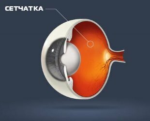
It is believed that such a visual impairment is associated with partial or complete exfoliation of the retina responsible for the projection of the visible image.
- 1. The main causes of development of the veil before the eyes and diseases in which this ailment occurs
- 2. Biological "welding" of the retina
- 3. Diagnosis of the veil before the eyes
- 4. Treatment of
- 5. Prevention of
In connection withthis, any deformations or changes in the retina reduce the visual acuity or even complete blindness of the person.
The main reasons for the development of the veil before the eyes and the diseases in which this ailment occurs
A shroud before the eyes can trigger the following situations and diseases.
- Diseases of the vessels .As a rule, with such ailments, complaints of a veil before the eyes are of an unstable nature and are often associated with impaired blood supply to the retina. Deterioration of vision occurs with the development of diseases such as hypotension, hypertension, anemia, vascular angiospasm of the retina, vegetative-vascular dystonia. In addition to the veils before the eyes, these diseases manifest themselves as a headache, a general weakness. Eliminating the cause of the shroud, the condition is normalized, however, in such cases, the patient will not need special ophthalmological care.
- Development of cataract .As a consequence of this disease, misting gradually increases, often for several years. This is due to the opacity of the biological lens - the lens. Therapy in this case consists in the use of special drops for the eyes with vitamins( Katachrom, Quinaks, Taufon, Katalin, etc.), but they can slow down the formation of the veil before your eyes, and not get rid of it. Unfortunately, without surgery in this case can not do - without fail it is necessary to replace the opacified lens with an artificial lens.
- Attack of glaucoma .Complaints on the shroud before the eyes appear sharply, and often this condition is accompanied by pain in the eye and a headache in the side of the lesion, as well as rainbow circles. In this situation, urgent help is needed from the ophthalmologist, inside you need to take a diuretic( a diuretic - furosemide) and drip into the eye of Pilocarpine, take an analgesic. In the absence of a positive effect, the patient is shown surgical treatment.
- Disrupted patency of retinal vessels .Such symptomatology grows quite quickly, right up to complete blindness. This condition develops against the background of the following diseases: atherosclerosis, hypertension, diabetes, etc. Depending on the venous thrombosis of the retina or embolism of the arteries, therapy is prescribed exclusively in the conditions of an ophthalmologic hospital.
- Diseases of the cornea of the eye .The cornea is a light-refracting and light-transmitting system of the eye, which, with the development of pathologies, loses its transparency and the light rays cease to fall on the retina of the eye. This process often proceeds with lightning speed, the treatment is determined only by the ophthalmologist.

Among the cause-effect relationships of the appearance of the shroud before the eyes are:
- Diabetes mellitus .This disease is one of the strongest provocators of retinal detachment. With the development of this ailment, very fragile blood vessels grow in the patient's eye, they poorly nourish the retina and easily burst. Such symptoms often lead to tissue dystrophy, and the blood that flows out of the bursted vessel can provoke retinal detachment.
- Retinal dystrophy .This is a common cause of retinal detachment, can occur with eye injuries, adrenal disease, rheumatoid arthritis, abnormalities in the thyroid gland and other diseases.
The retina of the eye becomes thinner in two cases:
- in the development of myopia ( as this changes the shape of the eyeball, which causes the retina to expand and deform from within),
- with insufficient supply of eye tissues( some diseases of the cardiovascular blood flow lead to the fact thatblood does not go to the organs of vision).
In those places where the retina is thinning, tears and cracks appear, the blood or vitreous body of the eye flows in them( a gel-like substance that fills the eye).
Moisture seeps through the cracks of the retina and lifts it - this is how detachment occurs. At the beginning of the detachment, the patient does not feel any discomfort, so it is necessary to visit an ophthalmologist once a year for advice.
Biological "welding" of the retina
So doctors call a painless procedure in which a laser beam points to the retina and how to "weld" the exfoliated tissues. The liquid that accumulates under the eye tissues dissipates after some time after the operation. This operation lasts from 30 minutes to 1 hour in a hospital. In 80% of cases after such intervention vision is restored by 98%.
It must be remembered that detachment of the retina is a disease that progresses and if it is ignored, then complete blindness will necessarily come.
Diagnosis of the veil before the eyes
Diagnosis for eyesight hazards is performed using the following tests:
- ophthalmological examination of a specialist for determining the retina,
- eye tonometry( or measurement of intraocular pressure),
- ultrasound,
- biomicroscopy( slit lamp examination).
Treatment of

Treatment of the veil before the eyes directly depends on the underlying disease, which causes turbidity. Let's consider some ways of treatment.
- In case of extensive damage to the cornea of the eye with scar tissue, the patient may be shown to have her transplant.
- When cataracts are effective, removal of the lens of the eye and implantation of the intraocular optical lens.
- In acute glaucoma, the patient is treated as an emergency, because there is a risk of loss of vision.
- At the initial stage of the cataract, in order to slow the pathological process, vitamins are buried in the diseased eye, and in the case of mature cataracts, only surgical treatment is indicated.
- The affected retina is treated with metabolic and vascular therapy, and laser coagulation is carried out, which helps prevent retinal exfoliation.
Prevention
Prevention of opacity in the eyes is aimed at preventing possible causes of its appearance. In this regard, the main recommendations are as follows:
- constant measurement of blood pressure level,
- constant monitoring of blood sugar level( to exclude vascular injury of the retina),
- hygiene( to prevent infection of the visual organs),
- after 40-it is recommended that the ophthalmologist be constantly and regularly monitored( it is at adulthood that the risk of glaucoma and cataracts increases).
It should also be noted that the symptom of the shroud before the eyes as an ophthalmic disease can occur both in the case of damage to the light-conducting and in the defeat of the light-receiving department of the visual organ.
Treatment is performed only after establishing a complete and accurate picture of the disease with the help of diagnostics, and afterwards both conservative and surgical treatment is prescribed.
